Category Archives: Thorax

ED Thoracotomy: Practice Management Guideline

I still encounter a quite a bit of confusion about ED thoracotomy: when to do it, when to stop. A nice set of guidelines was developed by the Subcommittee on Outcomes of the American College of Surgeons Committee on Trauma about 10 years ago. And really, things haven’t changed very much since then.

In general, the literature on this topic has been all over the place. Most studies are retrospective with very little statistical validation. But there’s a lot of it out there. The subcommittee used fairly standard methods to evaluate the literature and come up with some recommendations.

The overall survival rate for all comers was 8% (11% for penetrating, 1.6% for blunt). About 15% of survivors (no breakdown of blunt vs penetrating) suffered from neurologic impairment. Penetrating cardiac injury had the best survival (31%). In the 4 studies on children, the survival numbers paralleled the adults.

Because of the relatively weak quality of the data, only level II recommendations were given. They were:

  • Don’t consider ED thoracotomy in blunt trauma patients unless the patient arrests in front of you. Otherwise, meaningful survival is almost nonexistent.
  • Consider ED thoracotomy for penetrating chest injury if there were witnessed signs of life and a short transport time (5-10 minutes max, in my opinion). Look for pupillary response, spontaneous respirations, palpable pulse, or a narrow complex cardiac rhythm.
  • ED thoracotomy for penetrating abdominal injuries has a low survival rate, but can be considered if the injury may involve the chest.
  • Consider thoracotomy for suspected abdominal vascular injury, but survival is also very low.
  • The guidelines above apply equally to children.

Practical tips: ED thoracotomy is a seldom used and dangerous procedure. There are many opportunities for injury to trauma professionals, so be selective and take precautions. Assign someone to run the chemical code while the surgeons open the chest. Watch out for broken ribs and scalpels gone wild!

I’ll post pictures and specific pointers over the next three days.

Related posts:

Reference: Practice management guidelines for emergency department thoracotomy. JACS 193(3):303-309, 2001.

Fixation of Rib Fractures

Yesterday, I wrote about nonoperative management of rib fractures. Currently, the majority of rib fractures in this country are managed this way. During the past decade, a number of surgical rib fixation systems have been developed. The question is, when do you really need to consider this more invasive and potentially costly intervention? A review article from this hospital published earlier this year digs into the subsets of patients for whom operative management.

The Eastern Association for the Surgery of Trauma recently surveyed their own members, members of the Orthopedic Trauma Association, and a number of thoracic surgeons on the topic of operative rib fracture management. About 75% believed that operative fixation was indicated in some patients, but only about 20% or trauma surgeons and orthopedic surgeons and about half of thoracic surgeons had actually performed it.

The proposed benefits of surgical fixation are faster return of pulmonary function, fewer complications due to shorter ventilator time, shorter ICU and hospital lengths of stay, and a faster return to work. This review article found that these benefits were real when the technique is applied to select patients.

The authors found that:

  • The best indication is flail chest and respiratory failure without pulmonary contusion
  • Non-intubated patients with flail chest and deteriorating pulmonary function are also candidates
  • Reduction of pain and disability from symptomatic malunion or nonunion is a weaker indication due to sparse literature support
  • Other factors such as acute pain, open fractures, fracture repair while performing a thoracotomy for other reasons and chest wall deformity are weakly supported by the literature at best
  • There is no clear winner in the battle of hardware fixation systems

Bottom line: Operative rib fixation is indicated in patients with flail chest and pulmonary problems without significant pulmonary contusion, and in those with symptomatic mal- and non-unions. Flail chest patients benefit from early fixation, while the mal/nonunion groups should have fixation later once this condition is identified. Consideration for other indications should carefully take into account the cost, risk, and benefit to the patient. The literature is very weak in this regard, and a great deal more work is necessary to ensure that these techniques are not overused. 

Reference: Operative treatment of chest wall injuries: indications, technique, and outcomes. JBJS 93:97-110, 2011. 

When To Remove a Chest Tube

Chest tubes are needed occasionally to help manage chest injuries. How do you decide when they are ready for removal?

Unfortunately, the literature is not very helpful in answering this question. To come up with a uniform way of pulling them, our group looked at any existing literature and then filled in the blanks, negotiating criteria that we could all live with. We came up with the following.

Removal criteria:

  • No (or a minimal, stable) residual pneumothorax
  • No air leak
  • Less than 150cc drainage over the past 3 shifts. We do not use daily numbers, as it may delay the removal sequence. We have moved away from the “only pull tubes on the day shift” mentality. Once the criteria are met, we begin the removal sequence, even in the evening or at night.

Removal sequence:

  • Has the patient ever had an air leak? If so, they are placed on water seal for 6 hours and a followup AP or PA view chest x-ray is obtained. If no pneumothorax is seen, proceed to the next step.
  • Pull the tube. Click here to see a video demonstrating the proper technique.
  • Obtain a followup AP or PA view chest x-ray in 6 hours.
  • If no recurrent pneumothorax, send the patient home! (if appropriate)

Click here to download the full printed protocol.

Off-Label Use of the Foley Catheter

Foley catheters are a mainstay of medical care in patients who need control or measurement of urine output. Leave it to trauma surgeons to find warped, new ways to use them!

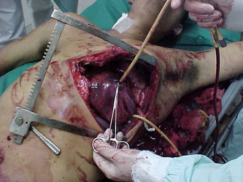
Use of these catheters to tamponade penetrating cardiac injuries has been recognized for decades (see picture, 2 holes!). Less well appreciated is their use to stop bleeding from other penetrating wounds.

foleyinheart

Foley catheters can be inserted into just about any small penetrating wound with bleeding that does not respond to direct pressure. (Remember, direct pressure is applied by one or two fingers only, with no flat dressings underneath to diffuse the pressure). Arterial bleeding, venous bleeding or both can be controlled with this technique.

In general, the largest catheter with the largest possible balloon should be selected. It is then inserted directly into the wound until the entire balloon is inside the body. Inflate the balloon using saline until firm resistance is encounted, and the bleeding hopefully stops. Important: be sure to clamp the end of the catheter so the bleeding doesn’t find the easy way out!

Use of catheter tamponade buys some time, but these patients need to be in the OR. In general, once other life threatening issues are dealt with in the resuscitation room, the patient should be moved directly to the operating room. In rare cases, an angiogram may be needed to help determine the type of repair. However, in the vast majority of cases, the surgeon will know exactly where the injury is and further study is not needed. The catheter is then prepped along with most of the patient so that the operative repair can be completed.

Related posts: