Category Archives: Head

Best Of EAST #10: (F)utility Of ICP Monitoring In Geriatric Patients?

Patients with severe TBI are typically managed using staged protocols based on the Brain Trauma Foundation (BTF) guidelines for ICP monitoring. There have been a number of papers over the past six years that question the utility of ICP monitoring, at least using the procedures in the BTF guidelines.  Most of these studies do not specifically break out elderly patients.

The group at the Westchester Medical Center in NY used the TQIP database to review the impact of ICP monitoring for severe TBI in patients > 65 years old. They performed a four year database study on these patients with an isolated head injury (no other body regions with AIS > 2), initial GCS < 8, and a length of stay > 24 hours. The examined the presence or absence of an ICP monitor, AIS head score, GCS, and a number of outcome measures.

Here are the factoids:

  • A total of 4,433 patients met the above criteria, and 17% had an ICP monitor placed
  • After propensity matching for those with and without an ICP monitor, mortality was nearly identical in both groups at 49%
  • ICU length of stay, hospital length of stay, and ventilator days were significantly longer in the monitor group

The authors concluded that ICP monitoring in this elderly group of patients did not improve survival and increased length of time in the ICU, hospital, and on the ventilator. The recommend that the current guidelines be improved to recognize these facts.

Bottom line: This is a nice, simple study that sought to answer just a few nice, simple questions. The mortality results are convincingly equal between the groups with and without an ICP monitor. The lengths of stay and ventilator days are statistically significantly longer with p values < 0.001. However, the actual numbers are not provided. I have seen many studies where statistically different numbers are not clinically relevant.

There are a number of papers that have come to similar conclusion on other or broader groups of TBI patients. Although we have specific guidelines on who gets a monitor and what we do with the numbers, there is growing doubt that their use actually helps. Perhaps it is time for us to review the data and make appropriate revisions!

Here are my questions for the authors and presenter:

  • Tell us about your propensity score matching. This will help us understand how similar the patient groups really were, with the exception of their ICP monitors.
  • Please provide the actual numbers for your lengths of stay and ventilator days. We need to be sure these are clinically and/or financially significant.
  • Have the results of this work prompted you to rework your own practice guidelines for treatment of severe TBI? I’m always interested if the group feels strongly enough about their work that they would consider changing their practice based on it.

Reference: ROLE OF ICP MONITORING IN GERIATRIC TRAUMA PATIENTS. EAST 35th ASA, oral abstract #33.


Print Friendly, PDF & Email

Best Of EAST #9: Routine Repeat Head CT For TBI Patients On Antithrombotic Agents

The data we use as guidance for repeat head CT in elderly patients who sustain mild TBI while taking antithrombotic therapy remains limited. There is a slowly growing consensus that the need is limited, but there is still a very wide variation in practice patterns.

The group at HCA Healthcare Nashville collected data from 24 system hospitals on this very specific cohort of patients: elderly (age > 55), head trauma with GCS 14-15, an initial head CT, and no other injuries with AIS > 2. They divided these patients into two groups based on whether they were currently taking antithrombotic (AT) therapy. Rate of delayed intracranial hemorrhage (ICH), need for neurosurgical intervention, and mortality were compared.

Here are the factoids:

  • About 3,000 patients were enrolled and only 10% had a repeat head CT
  • Of those who were rescanned, 10% of patients on meds had a new ICH vs 6% in those not taking meds (not statistically significant)
  • Extrapolating those numbers to all patients, the rate of delayed ICH would be 0.7% in patients not taking AT vs 1.0% for those who were (also not significant)
  • Mortality attributable to a head bleed occurred in only one patient who was made comfort care
  • There were no neurosurgical procedures performed in either group

The authors concluded that this specific subset of patients has a very low rate of delayed ICH, and that there are minimal clinical consequences in those that do. They do not support repeat head CT.

Bottom line: This abstract adds to the growing body of literature that shows little benefit to repeat head CT scan after a negative initial study, even if the patient is on blood thinners. Many previous studies involve only a single center and/or have smaller numbers. This one is larger because of the size of the HCA trauma system, and answers a simple set of questions on a limited subgroup of patients: elderly, mild TBI, with limited other injuries.

My back of the envelope power calculations show the authors may be a little short of the number of subjects to be able to show that the difference in the number of delayed ICH (0.7% vs 1.0%) is statistically significant. But the numbers are close enough and the p value so large (0.3) that they are probably right. This is completely offset by the absence of necessary neurosurgical interventions and the single attributable death.

Many trauma centers, including my own, have adopted a “no repeat scan” policy after a negative initial scan, even on thinners. In fact, unless the patient has some other injury that requires admission, they are discharged home with a responsible adult.

Here are my questions for the authors and presenter:

  • Did you do any type of power analysis to determine if the large number of patients included was actually large enough?
  • The term “antithrombotic therapy” is used broadly; which agents were considered in this category? Traditional warfarin therapy? Aspirin and other antiplatelet agents? DOACS?
  • Have you changed your system guidelines to reflect your work?

This is important and practical work! I’m looking forward to hearing all the details.

Reference: ROUTINE REPEAT BRAIN CT SCANNING IS UNNECESSARY IN OLDER PATIENTS WITH GCS 14-15 AND A NORMAL INITIAL BRAIN CT SCAN REGARDLESS OF PREINJURY ANTITHROMBOTIC USE: A MULTICENTER STUDY OF 3033 PATIENTS. EAST 35th ASA, oral abstract #31.

Print Friendly, PDF & Email

The Second Head CT In Patients Taking DOACs

Direct oral anticoagulant drugs (DOACs) are here to stay. When they were first released, I was very concerned with our inability to reverse them. I feared that we would have a rash of our elders presenting with severe head bleeds that we could do nothing about.

Well, that has not materialized. In fact, it appears that the probability of serious bleeding is more likely with our old reversible workhorse drug, warfarin.

But we are still spooked by DOACs. Nearly every center that has a practice guideline for managing patients with TBI on blood thinners includes a repeat CT scan after a given time interval. This is typically 6, 12, or even 24 hours.

Given the evolving safety profile of DOACs, is this even necessary? The surgical group at the Henry Ford Wyandotte Hospital in Michigan performed a retrospective registry review for their Level III trauma center. They reviewed the data for all adult patients who had suspected or confirmed blunt head trauma (any mechanism), were taking a DOAC, and received at least one CT scan.

Here are the factoids:

  • There were 400 patients with 498 encounters (yes, 15% came back with another TBI)
  • Patients were elderly (mean age 76) and nearly evenly split by sex
  • Fall was the most common mechanism (97%)
  • The first scan was negative in 96% of patients;12% of them did not have a repeat scan
  • Of the 420 patients who had a second scan, 418 were negative (99.5%). The two with positive scans were discharged uneventfully.
  • There were no differences based on specific DOAC, presenting GCS or mechanism

Bottom line: This is a relatively small, single institution study. However, it does appear that the authors have a large population of elderly patients suffering falls. This paper suggests that, indeed, a second scan may not be necessary. This parallels data from my own hospital. But to be on the safe side, keep an eye out for bigger, multi-institutional studies to be sure.

Reference: The utility of a second head CT scan af-ter a negative initial CT scan in head trauma patients on new direct oral anticoagulants (DOACs). Injury, article in press, June 13, 2021.

Print Friendly, PDF & Email

Hitting The Brakes May Increase Intracranial Pressure During The Ambulance Ride!

One of the most common injuries encountered by trauma professionals is blunt head trauma, and it’s one of the leading causes of death in young people. Keeping the level of intracranial pressure (ICP) within a specified range is one of the basic tenets of critical neurotrauma care in these patients. Most trauma centers have sophisticated algorithms that provide treatment guidance for various levels of ICP or cerebral perfusion.

The vast majority of patients with severe head injuries are transported to the hospital in some type of ambulance. Obviously, the exact ICP level is not known during transport because no monitoring device is present. We can sometimes infer that ICP is elevated if the patient has a Cushing response (wide pulse pressure and bradycardia) or unequal pupils. But for the most part, we assume that ICP is in a steady state during the ambulance ride.

But here’s something I never considered before: can ambulance acceleration or deceleration change the ICP through shifting of the brain or cerebrospinal fluid?

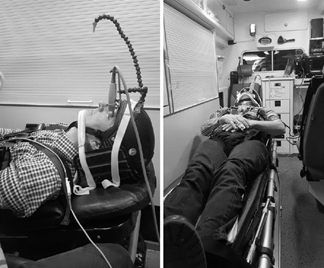
Patients are generally loaded into ambulances head-first, with their feet toward the back door. Frequently, they must be positioned supine in consideration of possible thoracic or lumbar spine injury. This position itself may lead to an increase in ICP. But what happens when the ambulance is hitting the brakes as it approaches a light or stop sign? As the patient’s weight shifts toward the top of the head, so does the CSF, spinal cord, and brain. Couldn’t this, too, increase ICP?

The anesthesiology group at the Erasmus Medical Center in Rotterdam, Holland performed a very novel study to assess this very thing. They recruited twenty participants in whom they evaluated ICP in various positions during acceleration and deceleration.
No, the subjects did not have an actual invasive ICP monitor inserted.

The authors used a novel way to infer pressures: optic nerve sheath diameter (ONSD). The optic nerves are direct extensions of the brain, and CSF travels freely in the nerve sheath. As ICP rises, the diameter of the nerve sheath increases. The subjects were fitted with a special helmet with two devices mounted on it. The first was a 7.5 Mhz ultra-sound probe focused on the back of the eye. The second was an arm with an orange dot on the end. This was adjusted so that the ultrasound probe was pointing at the optic nerve sheath when the other eye was focused on the dot. Subjects just watched the dot and measurements streamed in! Crude but very effective.

Baseline measurements were taken without acceleration or deceleration, then repeated when accelerating to 50 km/hr and decelerating to a stop.

Here are the factoids:

  • A total of 20 subjects were tested, and their oxygen saturation, blood pressure, and pulse were identical pre- and post-test
  • Baseline ONSD was about 5mm; a relevant change in diameter was determined to be more than 0.2 mm
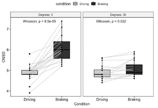
  • Lying supine, the ONSD in nearly all subjects increased from an average 4.8 to 6.0 mm during deceleration
  • With the head raised to 30º, most values remained steady (from 4.8 to 4.9 mm) during deceleration

The left block shows the increase in size of the optic disk with braking while supine. The right one demonstrates that this effect is neutralized by elevating the head 30º.

Bottom line: This is a small, simple, and creative study, yet the results are very interesting! It is clear that optic nerve sheath diameter increases significantly during deceleration in patients who are supine. And this effect is eliminated if the head of bed is elevated 30º.

Unfortunately, we have no idea how the change in ONSD corresponds to absolute values of, or relative increases in, ICP. Does a change of 1.2mm indicate a 5 torr increase in ICP? A 5% increase? Is it proportional to the absolute ICP? We just don’t know.

But the data is clear that a measurable change does occur. Until better data is available, it may be desirable to transport patients with serious head injuries with the head elevated to 30º if there are no concerns for lower spine injury. Or failing that, make sure the driver does not have a lead foot!

Reference: Ambulance deceleration causes increased intra cranial pressure in supine position: a prospective observational prove of principle study. Scand J Trauma Open Access 29:87, 2021.

Print Friendly, PDF & Email

Best Of AAST 2021: Validating The “Brain Injury Guidelines” (BIG)

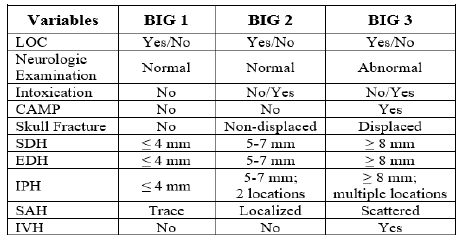
The Brain Injury  Guidelines (BIG) were developed to allow trauma programs to stratify head injuries in such a way as to better utilize resources such as hospital beds, CT scanning, and neurosurgical consultation. Injuries are stratified into three BIG categories, and management is based on it. Here is the stratification algorithm:

And here is the management algorithm based on the stratification above:

The AAST BIG Multi-Institutional Group set about validating this system to ensure that it was accurate and safe. They identified adult patients from nine high level trauma centers that had a positive initial head CT scan. They looked at the the need for neurosurgical intervention, change in neuro exam, progression on repeat head CT, any visits to the ED after discharge, and readmission for the injury within 30 days.

Here are the factoids:

  • About 2,000 patients were included in the study, with BIG1 = 15%, BIG2 = 15%, and BIG3 = 70% of patients
  • BIG1: no patients worsened, 1% had progression on CT, none required neurosurgical intervention, no readmits or ED visits
  • BIG2: 1% worsened clinically, 7% had progression on CT, none required neurosurgical intervention, no readmits or ED visits
  • All patients who required neurosurgical intervention were BIG3 (20% of patients)

The authors concluded that using the BIG criteria, CT scan use and neurosurgical consultation would have been decreased by 29%.

Bottom line: This is an exciting abstract! BIG has been around for awhile, and some centers have already started using it for planning the management of their TBI patients. This study provides some validation that the system works and keeps patients safe while being respectful of resource utilization. 

My only criticism is that the number of patients in the BIG1 and BIG2 categories is low (about 600 combined). Thus, our experience in these groups remains somewhat limited. However, the study is very promising, and more centers should consider adopting BIG to help them refine their management of TBI patients.

Reference: VALIDATING THE BRAIN INJURY GUIDELINES (BIG): RESULTS OF AN AAST PROSPECTIVE MULTI-INSTITUTIONAL TRIAL. AAST 2021, Oral abstract #25.

Print Friendly, PDF & Email