Category Archives: Head

Treatment Of BCVI

In my last post, I reviewed the grading system for blunt carotid and vertebral artery injury (BCVI). Today, we’ll discuss treatment, and in the next post, we will wrap up with pediatric-specific information.

There are basically three modalities at our disposal for managing BCVIantithrombotic medication (heparin and/or antiplatelet agents), surgery, and endovascular procedures. The choice of therapy is usually based on surgical accessibility and patient safety for anticoagulation. We know that several studies have shown decreased stroke events in heparinized patients. Unfortunately, this is not always possible due to associated injuries. Antiplatelet agents are usually tolerated after acute trauma, especially low-dose aspirin. However, several studies have shown little difference in outcomes in patients receiving heparin vs. aspirin/clopidogrel for BCVI.

So what to do? Here are some broad guidelines:

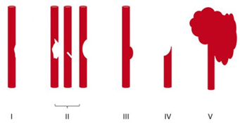
  • Grade I (intimal flap). Heparin or antiplatelet agents should be given. If heparin can be safely administered, it may be preferable in patients needing other surgical procedures since it can be rapidly reversed by stopping the infusion. These lesions generally heal entirely on their own, so a follow-up CT angiogram should be scheduled in 1-2 weeks. Medication can be stopped when the lesion heals.
  • Grade II (flap/dissection/hematoma). These injuries are more likely to progress, so heparin is preferred if it can be safely given. Stenting should be considered, especially if the lesion progresses. Long-term anti-platelet medication may be required.
  • Grade III (pseudoaneurysm). Initial heparin therapy is preferred unless contraindicated. Stable pseudoaneurysms should be followed with CTA every six months. If the lesion enlarges, surgical repair should be performed in accessible injuries or stenting in inaccessible ones.
  • Grade IV (occlusion). Heparin therapy should be initiated unless contraindicated. Patients who do not suffer a catastrophic stroke may do well with follow-up antithrombotic therapy. Endovascular treatment does not appear to be helpful.
  • Grade V (transection with extravasation). This lesion is frequently fatal, and the bleeding must be addressed using the best available technique. For lesions that are surgically accessible, the patient should undergo the appropriate vascular procedure. Inaccessible injuries should undergo angiographic treatment and may require embolization to control bleeding without regard for the possibility of stroke.

References:

  1. Scott WW, Sharp S, Figueroa SA, et al. Clinical and radiographic outcomes following traumatic Grade 1 and 2 carotid artery injuries: a 10-year retrospective analysis from a Level I trauma center. J Neurosurg 122:1196, 2015.
  2. Scott WW, Sharp S, Figueroa SA, et al. Clinical and radiographic outcomes following traumatic Grade 3 and 4 carotid artery injuries: a 10-year retrospective analysis from a Level 1 trauma center. J Neurosurg 122:610, 2015.
  3. Scott WW, Sharp S, Figueroa SA, et al. Clinical and radiological outcomes following traumatic Grade 1 and 2 vertebral artery injuries: a 10-year retrospective analysis from a Level 1 trauma center. J Neurosurg 121:450, 2015.
  4. Scott WW, Sharp S, Figueroa SA, et al. Clinical and radiological outcomes following traumatic Grade 3 and 4 vertebral artery injuries: a 10-year retrospective analysis from a Level I trauma center. The Parkland Carotid and Vertebral Artery Injury Survey. J Neurosurg 122:1202, 2015.

How To Grade BCVI

In my last post, I reviewed the three screening systems for blunt carotid and vertebral artery injury (BCVI). Today, we’ll look at grading them.

Just ten years ago, it was a major production to identify BCVI. Then, CT angiography was still in its early days, and scanner resolution and radiologist experience were significant issues that impacted accuracy.

We’ve come a long way in a relatively short period of time, and current-day scanners now have more than adequate resolution. It’s also more common to have a radiologist with special skills reading these studies, the neuroradiologist. For these reasons, CT angiography has become the standard for diagnosis. It is also the most cost-effective. Only in sporadic cases do we need to obtain a conventional contrast angiogram.

Once the study has been obtained, it’s time to identify and classify the injury. The Denver group is also responsible for bringing us the grading system for BCVI. See the diagram below.

Here are the details:

Grade I: A mild intimal irregularity is seen. Note the abnormally narrowed area, representing a minor intimal injury, possibly with a small amount of clot.

Grade II: This grade has several presentations. There may be an intraluminal thrombosis/hematoma with (left) or without (right) an intimal flap, or a flap alone (center).

Grade III: There is a full-thickness injury to the vessel with a contained extraluminal extravasation (pseudoaneurysm)

Grade IV: The vessel is completely occluded by a flap or thrombus

Grade V: The artery is transected and freely extravasating

Here’s a nice diagram:

Remember, we always grade things for a reason! Ultimately, the injury grade will translate into the selection of treatment. We’ll cover that in my next post.

Reference: Blunt carotid arterial injuries: implications of a new grading scale. J Trauma. 1999;47(5): 845-53.

How Common Is BCVI?

Blunt carotid and vertebral artery injuries (BCVI) are an under-appreciated problem after blunt trauma. Several screening tools have been published over the years, but they tend to be unevenly applied at individual trauma centers. I will discuss them in detail in the next section.

For the longest time, the overall incidence of BCVI was thought to be low, on the order of 1-2%. This is the number I learned years ago, and it has not really changed over time.

But how do we know for sure? Well, the group at Birmingham retrospectively reviewed every CT angiogram (CTA) of the neck they did in a recent two-year period. They did this after adopting a policy of imaging each and every one of their major blunt trauma patients for BCVI. Each patient chart was also evaluated to see if the patient met any of the criteria for the three commonly used screening systems.

During the study period, a total of 6,287 of 6,800 blunt trauma patients underwent BCVI screening with CTA of the neck. They discovered that 480 patients (7.6%) were positive for BCVI!

This is a shocking 8x higher than we expected! So why hasn’t this been obvious until now? Most likely because we were previously only aware of patients who became symptomatic. Luckily, many of these patients dodge the proverbial bullet and never exhibit any symptoms at all.

And what about pediatric patients? The neurosurgery groups at the University of New Mexico and Texas Children’s Hospital analyzed data in the Kids’ Inpatient Database (KID), which contains nationally representative pediatric data from the US. Five samples were obtained three years apart, beginning in 2000 and extending to 2012.

There were nearly 650,000 admissions for blunt trauma in the database, and 2150 were associated with BCVI. There was an interesting trend: incidence in 2000 started at 0.24% and increased to 0.49% in 2012. This represents a relative doubling of cases! Keep in mind that the absolute numbers remained very small, especially compared to the adult incidence.

Children aged 4 to 13 had the lowest risk of sustaining BCVI. This was higher in younger kids (ages 0-3), probably due to their big heads. It was also higher in adolescents and young adults (age 15-20). The injury was found more often in conjunction with cervical spine, skull base, clavicle, and facial fractures, as well as in children with TBI and intracranial hemorrhage.

Over one-third of children sustaining BCVI suffered a stroke (37%). Mortality was high, with a total mortality of 13%. This increased to a 20% rate if a stroke occurred.

So why should we be worried? This is one of those clinical entities like blunt thoracic aortic disruption that potentially has terrible consequences if ignored. And it seems to be worse among children even though it is far less common. Although the number of patients who develop sequelae from their BCVI is small, suffering a stroke can be catastrophic.

Should we perform a screening study for all blunt trauma patients? It seems like overkill, or is it? Is there any way we can be more selective about it?

In the next post, I’ll review the current screening tools used to determine which patients should receive CTA and how good they are.

References:

  1. Universal screening for blunt cerebrovascular injury. J Trauma 90(2):224-231, 2021.
  2. Blunt cerebrovascular injury in pediatric trauma: a national database study. J Neurosurg Pediatr. 2019

Subdural Hematomas and Hygromas Simplified

There’s a lot of confusion about subdural pathology after head trauma. All subdural collections are located under the dura, on the brain’s surface. In some way, they involve or can involve the bridging veins, which are somewhat fragile and get more so with age.

Head trauma causes a subdural hematoma by tearing some of these bridging veins. Notice how thick the dura is and how delicate the bridging veins are in the image below.

When these veins tear, bleeding ensues, which layers out over the surface of the brain in that area. If the bleeding does not stop, pressure builds and compresses and shifts the brain. A subdural hematoma is considered acute from the time of injury until about three days later. During this time, it appears more dense than brain tissue.

After about 3-7 days, the clot begins to liquefy and becomes less dense on CT. Many hematomas are reabsorbed, but occasionally there is repeated bleeding from the bridging veins, or the hematoma draws fluid into itself due to the concentration gradient. As a result, it can enlarge and begin to cause new symptoms. During this period, it is considered subacute.

It moves on to a more chronic stage over the ensuing weeks. The blood cells in it break down completely, and the fluid that is left is generally less dense than the brain underneath it. The image below shows a chronic subdural (arrows).

Hygromas are different because they are a collection of CSF, not blood. They are caused by a tear in the meninges and allow CSF to accumulate in the subdural space. This can also be caused by head trauma and is generally very slow to form. They can lead to slow neurologic deterioration and are often found on head CT in patients with a history of falls, sometimes in the distant past. CT appearance is similar to a chronic subdural, but the density is the same as CSF, so it should have the same appearance as the fluid in the ventricle on CT.

Best Of EAST 2023 #12: VTE Prophylaxis In Severe TBI

Time for another abstract on venous thromboembolic disease (VTE) prophylaxis, but this time in patients with severe head injury. VTE is a significant problem for trauma patients. Those with a potential source of bleeding from their injuries cause us to hesitate and consider the timing of chemical prophylaxis closely. Do we really want to cause more bleeding?

This is particularly problematic with intracranial hemorrhage, as the treatment is major brain surgery. Over recent years, the literature has been leaning toward earlier prophylaxis as soon as the intracranial blood has stopped evolving.

The EAST Multicenter Trials Group performed a seven-year retrospective review at 24 Level I and II trauma centers to assess the safety and efficacy of VTE chemoprophylaxis.  They divided patients into three groups: no prophylaxis, early prophylaxis (within 24 hours), and late prophylaxis (after 24 hours).

The authors assessed two endpoints: VTE occurrence and expansion of intracranial hemorrhage (ICH). They used several regression models to check their hypotheses.

Here are the factoids:

  • A total of 2,659 patients met the inclusion criteria. This averages out to 15 eligible patients per month per center. This is probably reasonable when combining a few high-volume centers with more lower volume centers.
  • Compared to early prophylaxis, patients who received late prophylaxis were twice as likely to develop VTE, although this was not statistically significant (p = 0.059)
  • Compared to early prophylaxis, patients who received no prophylaxis were a third less likely to develop VTE, although this, too, was not statistically significant (p = 0.39
  • About 25% of patients who received either early or late prophylaxis suffered an extension of their ICH, but only 17% of the no-prophylaxis group did
  • The regression model showed that the no prophylaxis group was 36% less likely to develop ICH extension compared to either early or late prophylaxis groups.

The workgroup concluded that the development of VTE was not dependent on the timing of the start of prophylaxis. Furthermore, patients who did not receive any prophylaxis had significantly decreased odds of ICH extension. The group recommended larger randomized studies to extend this work.

Bottom line: Shocker! This multicenter study suggests that the no prophylaxis and early prophylaxis groups had fewer VTE events than the late group, although these results were not statistically significant. This means that there wasn’t an advantage to giving the shot.

And the other major conclusion was that both early and late prophylaxis was associated with a significantly higher incidence of ICH extension. 

Roll these together, and you will find that neither early nor late prophylaxis help prevent VTE, yet they are both associated with additional bleeding in and around the brain! 

Heresy! I am trying to figure out what to make of these results. Perhaps the retrospective nature of the study and the wildcards this introduces influenced the results. It could be a study power problem, except the numbers were approaching significance that was unfavorable for prophylaxis.

I will be very interested to hear how the authors explain these findings. And yes, a well-powered randomized study would be great, but I don’t think many institutional review boards will be keen on a no-treatment group given our current fear of VTE. So don’t count on any real answers soon.

Reference: EARLY VTE PROPHYLAXIS IN SEVERE TRAUMATIC BRAIN INJURY: A PROPENSITY SCORE WEIGHTED EAST MULTICENTER TRIAL. EAST 2023 Podium paper #38.