Category Archives: Complications

Can Lead Poisoning Occur After A Gunshot?

This is a fairly common question from victims of gunshots and their families. As you know, bullets are routinely left in place unless they are superficial. It may cause more damage to try to extract one, especially if it has come to rest in a deep location. But is there danger in leaving the bullet alone?

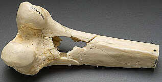
One of the classic papers on this topic was published in 1982 by Erwin Thal at Parkland Hospital in Dallas. The paper recounted a series of 16 patients who had developed signs and symptoms of lead poisoning (plumbism) after a gunshot or shotgun injury. The common thread in these cases was that the injury involved a joint or bursa near a joint. In some cases the missile passed through the joint/bursa but came to rest nearby, and a synovial pseudocyst formed which included the piece of lead. The joint fluid bathing the projectile caused lead to leach into the circulation.

The patients in the Parkland paper developed symptoms anywhere from 3 days to 40 years after injury. As is the case with plumbism, symptoms were variable and nonspecific. Patients presented with abdominal pain, anemia, cognitive problems, renal dysfunction and seizures to name a few.

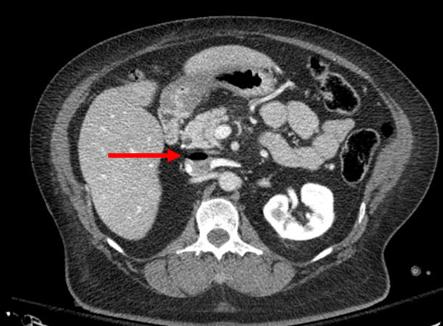
Bottom line: Any patient with a bullet or lead shot that is located in or near a joint or bursa should have the missile(s) promptly and surgically removed. Any lead that has come to rest within the GI tract (particularly the stomach) must be removed as well. If a patient presents with odd symptoms and has a history of a retained bullet, obtain a toxicology consult and begin a workup for lead poisoning. If levels are elevated, the missile must be extracted. Chelation therapy should be started preop because manipulation of the site may further increase lead levels. The missile and any stained tissues or pseudocyst must be removed in their entirety.

Granted, this is a very old paper. Over the years, a few papers on the topic have popped up from time to time. In my next post, I’ll review a meta-analysis on this topic that was published just last year.

Reference: Lead poisoning from retained bullets. Ann Surg 195(3):305-313, 1982.

What Does A Retained Surgical Sponge Look Like?

Surgeons and surgical residents rarely see these. And because it’s so uncommon, they frequently don’t recognize the telltale findings on radiographic studies. The TSA runs into the same problem in screening passengers for weapons and other hazards at airports. But it’s the bane of any surgeon’s existence. And it’s a major reason why OR personnel take such great pains to account for everything in the room. It is a catastrophe, and always a preventable one, when some piece of equipment goes missing and ends up left inside a patient.

A number of methods have been developed to try to eliminate this problem. They include careful counts, having someone record anytime anything is placed inside, x-rays, and most recently, RFID tags.

After counting, x-ray is the most common way to try to find missing objects. One would think that these foreign bodies would be easy to see. Metallic instruments are rather easy to spot. But many trauma professionals, even those who work in the OR, have never seen what a positive image of a sponge actually looks like. So here they are. You should never miss one on an xray now.

Surgeons typically use two types of sponges in the OR: Ray-Tec sponges and standard lap pads. Ray-Tecs look like a 4×8 piece of gauze with a mysterious blue string woven throughout it. The string is the only part that shows up on x-ray, and it is very thin and somewhat hard to see. Here are some Ray-Tec sponges outside the body:

And here’s one that was left inside. Note the little squiggle in the left lower quadrant and how easy it is to overlook.

On the other hand, a laparotomy pad is a 4×4 folded cloth pad that unfolds into a larger pad. It has a blue radiopaque tag sewn in the corner, extending along one edge of the pad. Here’s what they look like outside the body:

And here’s one inside a patient. Note the irregular object in the right upper quadrant. Many times the tag is scrunched up and doesn’t look like one.

Bottom line: It’s important for anyone who works in the OR on any body part to be familiar with the appearance of these tags on x-rays. Since it’s generally impossible to get accurate counts before or after a trauma procedure, always image the involved body cavity looking for these telltale signs before closing the patient.

Note: These images  were taken from the internet. Patients were not treated at Regions Hospital.

Air Embolism From an Intraosseous (IO) Line

Intraosseous (IO) lines are a godsend when we are faced with a patient who desperately needs access but has no veins. The tibia is generally easy to locate and the landmarks for insertion are straightforward. They are so easy to insert and use, we sometimes “set it and forget it”, in the words of infomercial guru Ron Popeil.

But complications are possible. The most common is an insertion “miss”, where the fluid then infuses into the knee joint or soft tissues of the leg. Problems can also arise when the tibia is fractured, leading to leakage into the soft tissues. Infection is extremely rare.

This photo shows the inferior vena cava of a patient with bilateral IO line insertions (black bubble at the top of the round IVC).

During transport, one line was inadvertently disconnected and probably entrained some air. There was no adverse clinical effect, but if the problem is not recognized and the line is not closed properly, there could be.

Bottom line: Treat an IO line as carefully as you would a regular IV. You can give anything through it that can be given via a regular IV: crystalloid, blood, drugs. And even air, so be careful!

Best Of EAST #9: Is TXA Associated With VTE?

Most trauma programs can be divided into two types: those that believe in tranexamic acid (TXA) and those that don’t. I won’t get into the details of the CRASH-2 study here. But those centers that don’t believe usually give one of two reasons: they don’t think it works or they think the risk of venous thromboembolism (VTE) is too high.

EAST put together a multi-institutional trial to see if there was an association between TXA administration and subsequent VTE. The results are being reported as one of the paper presentations at the meeting this week. A retrospective study of the experience of 15 trauma centers was organized. A power analysis was preformed in advance, which showed that at least 830 patients were needed to detect a positive result.

Adult patients who received more than 5 units of blood during the first 24 hours were included. There were a lot of exclusionary criteria. They included death within 24 hours, pregnancy, pre-injury use of anticoagulants, interhospital transfer, TXA administration after 3 hours, and asymptomatic patients that had duplex VTE surveillance (huh?). The primary outcome studied was incidence of VTE, and secondary outcomes were MI, stroke, length of stay, and death.

Here are the factoids:

  • There were 1,333 eligible patients identified, and 887 (67%) received TXA
  • Females were significantly (over 2x) more likely to receive TXA (46% vs 19%)
  • 80% of patients given TXA received VTE prophylaxis, whereas only 60% of those who did not receive TXA got prophylaxis (also significant)
  • TXA patients had a statistically significantly higher ISS (27) compared to non-TXA patients (25) but this is not clinically significant
  • Mortality in the TXA group was significantly lower (17% vs 34%)
  • The number of units of blood, plasma, and platelets transfused were significantly lower in the TXA group
  • VTE rate appeared lower in the TXA group, but once multivariate analysis was applied, there was no difference

The group concluded that there was no association between TXA and VTE, but that it was linked to decreased mortality and transfusion need.

My comment: This was a study done the way they are supposed to be! Know your objectives and study outcomes up front. Figure out how many patients are needed to tease out any differences. And use understandable statistics to do so.

But, of course, it’s not perfect. No retrospective study is. Nor is any multi-institutional trial. There are lots of little variations and biases that can creep in. But the larger than required sample size helps with reducing the noise from these issues.

Basically, we have a decent study that shows that the clinical end points that we usually strive for are significantly improved in patients who have TXA administered. We don’t know why, we just know that it’s a pretty good association.

This study shows that the usual reasons given for not using TXA don’t appear to hold true. So hopefully it will convert a few of the TXA non-believers out there. I’m excited to hear more details during the presentation at the meeting.

Reference: Association of TXA with venous thromboembolism in bleeding trauma patients: an EAST multicenter study. EAST Annual Assembly, Paper #13, 2020.

Best Of EAST #8: Early vs Late Full Anticoagulation In TBI

Trauma professionals are always reluctant to anticoagulate TBI patients with demonstrated blood in their head. In recent years, we’ve become more comfortable providing prophylactic doses of low molecular weight heparin after a suitable period. This is typically 24-48 hours after a stable head CT in patients with select types of intracranial hemorrhage (ICH) who are at increased risk for venous thromboembolism.

But what about therapeutic dose anticoagulation in these patients? Let’s say that you have a patient with ICH who has developed a significant pulmonary embolism (PE)? Is is safe to give full dose anticoagulation? And if so, when?

The group at Shock Trauma in Baltimore attempted to answer this in one of the EAST Quick Shot presentations scheduled for this week. The did a retrospective review of 4.5 years of their own data on these patients. They specifically selected patients who had both ICH and PE and compared those who received full anticoagulation within 7 days of injury vs those who were dosed after 7 days. Outcomes studied included death, interventions for worsening ICH, and pulmonary complications.

Here are the factoids:

  • A total of 50 patients had both ICH and PE, but only the 46 who received therapeutic anticoagulation were analyzed
  • 19 patients (41%) received early anticoagulation, and 27 received it late (59%)
  • There were 4 deaths in the early group (2 from the PE, 1 from multi-system organ failure, 1 from the TBI) vs none in the late group, and this was statistically significant
  • 3 patients in the early group (18%) vs 2 in the late group (7%) had an increase in their ICH (p=0.3), and none required intervention

The authors concluded that their study failed to show any instances of clinically significant progression of ICH after anticoagulation, and that it is not associated with worse outcomes, even if started early. Thus they recommend that ICH should not preclude full anticoagulation, even early after injury.

My comment: I always say that you shouldn’t let one paper change your practice. Even a really good one. In order to ensure that you are providing the best care, more work must always be done to confirm (or refute) the findings of any provocative research. And this little Quick Shot, with little opportunity for questions from the audience, should definitely not change it!

The major issues to consider here are common ones: 

  • This was a retrospective study and it does not appear that any guideline was followed to determine who got early vs late anticoagulation. So who knows what kind of selection bias was occurring and how the surgeon decided to prescribe anticoagulation? It’s very possible that patients with a “bad CT” were put into the late group, and the not so bad ones in the early group. This would bias the results toward better outcomes in the early anticoagulation group.
  • It’s also a very small study that is extremely underpowered. The authors comment on the fact that the outcomes of the early group were not worse than the late group. However, looking at their sample size (46) shows that they would only be able to show differences if they were about 5x worse in the early group. They would realistically need about 350 total patients to truly show that the groups behaved the same. Their small numbers also preclude saying that there were no ICH progressions. There very well could have been if 300 more patients were added to the series.
  • And isn’t death a significant outcome? The authors indicated that 2 of the 4 deaths were a result of the PE. Yet there was a significant association (p=0.02) of increased death in the early anticoagulation patients that can’t be discounted.

Bottom line: It’s way too early to consider giving early anticoagulation to patients with ICH and pulmonary embolism. It may very well be shown to be acceptable, eventually. But not yet. And a much bigger prospective study will be required to confirm it.

Reference: Therapeutic anticoagulation in patients with traumatic brain injuries and pulmonary emboli. EAST Annual Assembly Quick Shot #7, 2020.