Category Archives: Complications

More On Lead Poisoning And Retained Bullets

Trauma professionals frequently have to leave bullets in patients. It is often more disruptive to go digging the projectiles out than to just leave them in place. But patients always want to know why and what the consequences might be.

In my last post, I discussed a very old paper on what we know about lead levels and retained bullets. Very recently, a meta-analysis was published that provides a better picture of this topic. They somehow managed to find over 2000 articles dealing with lead toxicity and bullets out there. But after someone had the pleasure of reviewing each of them, they found only 12 that had any meaningful or actionable information.

Here are the factoids:

  • All studies were observational (duh! It would be difficult to get your IRB to approve a study where patients were shot on purpose)
  • There were five cross-sectional studies, four case-control studies, and three prospective cohort studies
  • The studies were small, with a median of only 26 patients (range 15-120)
  • Eleven of the twelve studies showed an association with retained bullets and elevated blood lead levels
  • Three studies showed elevated blood levels if a fracture was present
  • The higher the number of retained fragments, the more likely lead levels were to be high
  • Higher lead levels were associated with retained fragments near a bone or joint
  • There were no good correlations with number of fragments and location vs actual lead toxicity

Bottom line: Even using meta-analysis, it is difficult to tease out meaningful answers to this question. That speaks to the low numbers of papers and their quality. However, this study does provide a little bit of guidance.

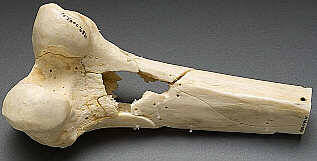
Retained bullet fragments are probably not a big worry in most patients. The bothersome cases are those where the fragments are in or near a bone or joint. And even though few patients actually developed lead toxicity, lead levels approaching 5 micrograms/dL can have physiologically significant negative effects. 

Recommendation: If your patient has a retained bullet fragment near a bone or joint, or they have “multiple” retained fragments (no good definition of this), they should have blood lead levels measured every three months for a year. If the level is rising, and certainly if it reaches the 5μ/dL level, attempts should be made to remove the fragments.

Reference: Lead toxicity from retained bullet fragments: A systematic review and meta-analysis. J Trauma 87(3):707-716, 2019.

Print Friendly, PDF & Email

Can Lead Poisoning Occur After A Gunshot?

This is a fairly common question from victims of gunshots and their families. As you know, bullets are routinely left in place unless they are superficial. It may cause more damage to try to extract one, especially if it has come to rest in a deep location. But is there danger in leaving the bullet alone?

One of the classic papers on this topic was published in 1982 by Erwin Thal at Parkland Hospital in Dallas. The paper recounted a series of 16 patients who had developed signs and symptoms of lead poisoning (plumbism) after a gunshot or shotgun injury. The common thread in these cases was that the injury involved a joint or bursa near a joint. In some cases the missile passed through the joint/bursa but came to rest nearby, and a synovial pseudocyst formed which included the piece of lead. The joint fluid bathing the projectile caused lead to leach into the circulation.

The patients in the Parkland paper developed symptoms anywhere from 3 days to 40 years after injury. As is the case with plumbism, symptoms were variable and nonspecific. Patients presented with abdominal pain, anemia, cognitive problems, renal dysfunction and seizures to name a few.

Bottom line: Any patient with a bullet or lead shot that is located in or near a joint or bursa should have the missile(s) promptly and surgically removed. Any lead that has come to rest within the GI tract (particularly the stomach) must be removed as well. If a patient presents with odd symptoms and has a history of a retained bullet, obtain a toxicology consult and begin a workup for lead poisoning. If levels are elevated, the missile must be extracted. Chelation therapy should be started preop because manipulation of the site may further increase lead levels. The missile and any stained tissues or pseudocyst must be removed in their entirety.

Granted, this is a very old paper. Over the years, a few papers on the topic have popped up from time to time. In my next post, I’ll review a meta-analysis on this topic that was just recently published.

Reference: Lead poisoning from retained bullets. Ann Surg 195(3):305-313, 1982.

Print Friendly, PDF & Email

AAST 2019 #6: DOACs Part 3!

A little further down the direct oral anticoagulants (DOACs) rabbit hole please? The abstract reviewed in my last post suggested that elderly patients taking these agents actually do better than those on warfarin. So if that’s the case, do we need to be so attentive to getting followup CT scans on these patients to ensure that nothing new and unexpected is happening?

The trauma group at UCSF – East Bay performed a multi-center review of the experience at “multiple” Level I trauma centers over a three year period. They included anticoagulated patients with blunt trauma who had a negative initial head CT. Patients taking only an anti-platelet agent or a non-oral anticoagulant were excluded.  They analyzed the data for new, delayed intracranial hemorrhage, use of reversal agents, neurosurgical intervention, readmission, and death.

Here are the factoids:

  • A total of 739 records were studied: 409 on warfarin and 330 on a DOAC. Average age was 79, and half were male.
  • Repeat head CT was performed only half the time (!)
  • Delayed hemorrhage was noted in 4% of warfarin cases (9 of 224) and 2.5% of DOAC cases (4 of 159)
  • There were no interventions or deaths in the DOAC group with followup CT, or in those who did not have the repeat scan
  • There was 1 intervention in the warfarin group and two deaths attributed to TBI
  • Reversal agents were administered to 2% of DOAC patients and 14% of warfarin patients
  • The authors performed a regression analysis that showed the two strong associations with delayed hemorrhage were male sex and AIS head > 2 (!)

The authors concluded that this “largest study” suggests that DOACs “may” have a better safety profile compared to warfarin and repeat head CT is not indicated.

Now, hold on a minute!

Rule #1: No single published paper should ever change your practice. They need to be confirmed by other, hopefully better work.

Rule #2: No single abstract should make you even think about changing your practice! These are preliminary works that always need more detail, more effort, and a lot more thought. They are meant to telegraph what the authors are working on and to raise interesting questions from the audience. They should stimulate others to try to replicate and improve upon the work. In general, if something looks really good as an abstract, the next step is successful publication. This means that peers have reviewed the data and agree that it looks promising. But then it should take several years of work by the original authors and others to prove or refute the claims.

This study was small in the first place, and became smaller because half did not have repeat CT scans. The only statistically significant result was that we confirmed that the providers were not very good about getting followup scans. Just because they didn’t do it doesn’t mean it’s not indicated, especially given the nature of the data and the very small numbers.

I consider this another very small piece in the puzzle that suggests DOACs are not as evil as warfarin. There are several of these low power studies floating around right now. But we need to hunker down and really do a big study right so we can start to get a clearer picture of what we should do. For now, it’s best to treat all anticoagulants and anti-platelet agents as evil and err on the side of overtreating.

Here are my comments and questions for the presenter and authors:

  • Why was the followup head CT rate so poor? Was this a “however they like to do it” thing, was there a protocol, did the trauma centers just not believe that DOACs could be bad?
  • What were the guidelines for reversal? If the initial head CT was normal, why ever reverse? This suggests that participating centers could do whatever they wanted based on unspecified criteria.
  • Was the regression analysis helpful in any way? Being male and having a mild TBI seem rather nonspecific factors and wouldn’t help select patients for reversal or repeat scan.
  • Please provide more information on the warfarin intervention and deaths.
  • Isn’t the title of this abstract rather bold for the quality of the results presented?

I’m sure there will be some lively debate at the end of this presentation!

Reference: Repeat CT head scan is not indicated in trauma patients taking novel anticoagulation: a multi-institutional study. AAST 2019, Oral Abstract #66.

Print Friendly, PDF & Email

AAST 2019 #5: DOACs Part 1

A short while ago I wrote about the proper nomenclature of the new or novel oral anticoagulant medications that are replacing warfarin in patients with atrial fibrillation (click here for details). Cut to the chase, the consensus seems to be that they should be called direct oral anticoagulants or DOACs.

These medications strike fear into the average trauma professional, primarily because there is no easy way to reverse them as there is for warfarin. We are finally accumulating enough experience with them to start to see the bigger picture with respect to complications and mortality. Today, I’ll begin the discussion with a series of three abstracts regarding these drugs.

The AAST conducted a multicenter, prospective, observational study that collected DOAC trauma patient information from 15 centers. They reviewed four years of data, specifically examining the use of reversal agents and mortality.

Here are the factoids:

  • A total of 606 patients were enrolled. They were generally elderly with an average age of 75.
  • Most were taking one of the Factor Xa inhibitors (apixaban, rivaroxaban, edoxiban), while just 8% were taking the direct thrombin inhibitor dabigatran.
  • Only 1% of patients received a reversal agent (prothrombin complex concentrate (PCC) 87%, Praxbind (12%), and Andexxa (1%)
  • Those receiving reversal tended to be older than the average and had more severe head injuries
  • Patients who were reversed with PCC had no change in mortality using a regression model
  • Patients reversed with Praxbind or Andexxa had a 15x higher probability of mortality

The author’s conclusions merely restated their results.

This is fascinating information. Unfortunately, this study was not designed to provide a comparison with patients taking warfarin. However, my next two abstract reviews will cover this very topic. 

There are two interesting tidbits here. First, reversal was only carried out in about one in eight patients. Why is this? No protocol? No product? Too pricey? Patients not hurt badly enough? And how would that be judged anyway?

The second is that reversal with PCC seems to be benign, but use of one of the specifically designed reversal agents really jacked up mortality. These agents (Praxbind and Andexxa) are very expensive ($3.5K and $50K respectively). Furthermore, there are no studies anywhere that show their effectiveness. This one actually seems to show they might be dangerous.

The devil is in the details. Here are my questions for the presenter and authors:

  • Were there any guidelines for reversal? This is key because if not, the statistics just describe “how we do it.” Yes, you can tease out higher ISS or AIS head as potential reasons, but were there directions regarding this built into the study protocol?
  • Do you have any data on the success rates of PCC reversal? Were there provisions to demonstrate lesion stability vs progression after administration?
  • Do you have an impression of why the tailored reversal agents seemed to be so deadly? Were they used as a last resort due to cost. Did the centers have a hard time getting it or authorizing its use?

This abstract could be a gold mine!

Reference: The AAST prospective, observational, multicenter study investigating the initial experience with reversal ofnovel oral anticoagulants in trauma patients. AAST 2019, Oral Paper 58.

Print Friendly, PDF & Email

AAST 2019 #2: Predicting Abdominal Operation After Blunt Trauma – The RAPTOR Score

Patients with blunt abdominal injury, particularly those with seat belt signs, can be diagnostically very challenging. If the patient is stable and does not have peritonitis, CT scan is typically the first stop after the trauma resuscitation room. As many trauma professionals know, the radiographic findings can be subtle and/or not very convincing.

The trauma group at the University of Tennessee in Memphis sought to identify specific findings that might help us better identify patients that will need laparotomy. They retrospectively identified all their mesenteric injuries over a five-year period. A single blinded radiologist (is this an oxymoron or not?) reviewed all 151 patient images who underwent laparotomy, looking for predictors of bowel or mesenteric injury.  All of the predictors were then converted into a scoring system called RAPTOR (radiographic predictors of therapeutic operative intervention; kind of a stretch?). These predictors were then subjected to multivariate regression analyses to try to tease out if there were any independent predictors of injury.

Here are the factoids:

  • A total of 151 patients were identified over the 5 year period; 114 underwent laparotomy
  • Of the 114 operated patients, two thirds underwent a therapeutic laparotomy and the other third were nontherapeutic
  • There no missed injuries in the non-operated patients
  • The components of the RAPTOR score were culled from all the potential findings, and were determined to be
    • Multifocal hematoma
    • Acute arterial extravasation
    • Bowel wall hematoma
    • Bowel devascularization
    • Fecalization (of what??)
    • Free air
    • Fat pad injury (??)
  • Linear regression then showed that only three of these, extravasation, bowel devascularization, and fat pad injury to be independent predictors of injury
  • If three or more RAPTOR variables were present, then the sensitivity, specificity, and positive predictive values for injury were 67%, 85%, and 86%, and an area under the receiver operating characteristic curve (AUROC) of 0.91

The authors concluded that the RAPTOR score provided a simplified approach to detect patients who might benefit from early laparotomy and not serial abdominal exams. They go further and say it could potentially be an invaluable tool when patients don’t have clear indications for operation.

It looks like there are two things going on here at the same time. First, a new potential scoring system is being piloted. And second, a regression analysis is being used to examine the data as well. 

But first, let’s back up to the beginning. This is a retrospective study, with a relatively small size. This makes it far harder to ensure that the results will be significant, or at least meaningful. Use of a single radiologist can also be problematic, especially since many of the CT findings with this mechanism of injury are subtle. 

The reported performance of the RAPTOR score is a bit weak. The listed statistics show that it accurately identified only two thirds of those who needed an operation and 85% of those who didn’t. The AUROC for the regression is very good, though. Could a good old-fashioned serial exam scenario be better?

Bottom line: It will be interesting to hear the background on RAPTOR vs regression, and find our how the authors will use or are using these tools.

Here are my questions for the presenter and authors:

  • Why did you decide to create a scoring system that uses a set of variables that may be dependent on each other? Isn’t the regression equation better?
  • Has this information changed your practice? It seems that the two of the three regression variables are fairly obvious reasons to operate (active extravasation and devascularization). Do you really need the rest?
  • Has this study helped you decrease the non-therapeutic laparotomy rate for blunt abdominal injury?
  • And please define fecalization and fat pad injury!

I’m looking forward to hearing this presentation!

Reference: RADIOGRAPHIC PREDICTORS OF THERAPEUTIC OPERATIVE INTERVENTION AFTER BLUNT ABDOMINAL TRAUMA: THE RAPTOR SCORE. AAST 2019 Oral Paper 6.


Print Friendly, PDF & Email