When To Obtain A Dedicated Facial CT

Initial CT scan evaluation for blunt trauma patients is fairly standardized. The usual palate consists of scans of the head, cervical spine, chest, abdomen and pelvis. Some choose their “colors” individually, and others just slop everything on the canvas.

However, there are a few other scans that are occasionally helpful and/or necessary. Think soft tissue views, or CT angiogram of the neck, or CT angiogram of potential extremity vascular injuries.

Another study that is occasionally needed but many times unnecessarily ordered is the dedicated CT of the facial bones. This study spans the entire area from mandible to frontal sinus and is performed using finer cuts to display greater detail.

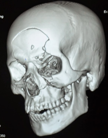
The unfortunate truth is that a large number of dedicated facial CTs either do not show fractures, or show fractures that don’t require operation. The scan does deliver a nice dose of radiation, though. Is there any way to be more selective about ordering it?

About 10 years ago, a plastic surgery group in Madison developed what came to be called the “Wisconsin criteria” for ordering facial imaging.  Here they are:

  • Bony step-off
  • Periorbital ecchymosis
  • GCS < 14
  • Malocclusion
  • Missing teeth

The authors claimed 97% sensitivity and 2.6% missed fracture rate, although external validation suggested those numbers were a bit generous. The Plastic Surgery group from the University of Minnesota and Regions Hospital recently re-studied these criteria with a large number of patients, looking at accuracy as well as cost-savings.

They performed a retrospective review of 1000 patients (based on a power analysis) who had a facial CT and adequate documentation of the Wisconsin criteria in the chart. Here are the factoids in table form:

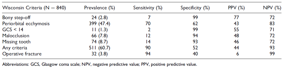
(click table for larger copy)

  • Periorbital ecchymosis was the most common criterion, which had the highest sensitivity of 70% (terrible)
  • The other criteria fared even worse from a sensitivity standpoint
  • But if you roll them all up together, the presence of any one of the five yielded a 90% sensitivity (true positives) and 52% specificity (true negatives)
  • The negative predictive value was 93% if none of the criteria were present, which means it’s a good tool for ruling out the need for a CT scan
  • The overall missed fracture rate was 2.8%, and only 0.12% for ones that required operation
  • Cost savings by limiting CT to patients who met the criteria was over $300K in 2014

Bottom line: What to do? It’s clear that using the absence of any of the Wisconsin criteria to avoid a facial CT scan is helpful. This makes sense, because 4 of the 5 criteria are findings on facial exam. But it also means that a lot of scans will still get done for low sensitivity criteria. 

How about this? Since nearly all of these patients will have head and cervical CT scans, review the head scan first for facial fractures. Single, non-displaced fractures are nearly always nonoperative in nature. If patterns of fractures are present, or there are significant displacements, a dedicated facial scan will be very helpful in determining operative management.

But remember, the head CT does not include the mandible. A good physical exam and occlusion check is mandatory, and any suspicion of injury should prompt a full scan of the face.

Thanks to Chris Stewart, the lead author on this study for sending it to me for review.

Rreference: Validation of the “Wisconsin criteria” for obtaining dedicated facial imaging and its financial impact at a Level I trauma center. Craniomaxillofacial Trauma & Recon 13(1):4-8, 2020.

The Trauma Activation Pat-Down?

Yes, this is another one of my pet peeves. During a trauma activation, we all strive to adhere to the Advanced Trauma Life Support protocols. Primary survey, secondary survey, etc. Usually, the primary survey is done well.

But then we get to the secondary survey, and things get sloppy.

police_frisk1

The secondary survey is supposed to be a quick yet thorough physical exam, both front and back. But all too often it’s quick, and not so thorough. There is the usual laying on of the hands, but barely. Abdominal palpation is usually done well. But little effort is put into checking stability of the pelvis. The extremities are gently patted down with the hope of finding fractures. Joints are slightly flexed, but not stressed at all.

Is it just a slow degradation of physical exam skills? Is it increasing (and misguided) faith in the utility of the CT scanner? I don’t really know. But it’s real!

Bottom line: Watch yourself and your team as they perform the secondary survey! Your goal is to find all the injuries you can before you go to imaging. This means deep palpation, twisting and trying to bend extremities looking for fractures, stressing joints looking for laxity. And doing a good neuro exam! Don’t let your physical exam skills atrophy! Your patients will thank you.

Serial Hemoglobin / Hematocrit – Huh? Part 2

In my last post, I waxed theoretical. I discussed the potential reasons for measuring serial hemoglobin or hematocrit levels, the limitations due to the rate of change of the values, and conjectured about how often they really should be drawn.

And now, how about something more practical? How about an some actual research? One of the more common situations for ordering serial hemoglobin draws occurs in managing solid organ injury. The vast majority of the practice guidelines I’ve seen call for repeating blood draws about every six hours. The trauma group at the University of Florida in Jacksonville decided to review their experience in patients with liver and spleen injuries. Their hypothesis was that hemodynamic changes would more likely change management than would lab value changes.

They performed a retrospective review of their experience with these patients over a one year period. Patients with higher grade solid organ injury (Grades III, IV, V), either isolated or in combination with other trauma, were included. Patients on anticoagulants or anti-platelet agents, as well as those who were hemodynamically unstable and were immediately operated on, were excluded.

Here are the factoids:

  • A total of 138 patients were included, and were separated into a group who required an urgent or unplanned intervention (35), and a group who did not (103)
  • The intervention group had a higher ISS (27 vs 22), and their solid organ injury was about 1.5 grades higher
  • Initial Hgb levels were the same for the two groups (13 for intervention group vs 12)
  • The number of blood draws was the same for the two groups (10 vs 9), as was the mean decrease in Hgb (3.7 vs 3.5 gm/dl)
  • Only the grade of spleen laceration predicted the need for an urgent procedure, not the decrease in Hgb

Bottom line: This is an elegant little study that examined the utility of serial hemoglobin draws on determining more aggressive interventions in solid organ injury patients. First, recognize that this is a single-institution, retrospective study. This just makes it a bit harder to get good results. But the authors took the time to do a power analysis, to ensure enough patients were enrolled so they could detect a 20% difference in their outcomes (intervention vs no intervention). 

Basically, they found that everyone’s Hgb started out about the same and drifted downwards to the same degree. But the group that required intervention was defined by the severity of the solid organ injury, not by any change in Hgb.

I’ve been preaching this concept for more than 20 years. I remember hovering over a patient with a high-grade spleen injury in whom I had just sent off the requisite q6 hour Hgb as he became hemodynamically unstable. Once I finished the laparotomy, I had a chance to pull up that result: 11gm/dl! 

Humans bleed whole blood. It takes a finite amount of time to pull fluid out of the interstitium to “refill the tank” and dilute out the Hgb value. For this reason, hemodynamics will always trump hemoglobin levels for making decisions regarding further intervention. So why get them?

Have a look at the Regions Hospital solid organ injury protocol using the link below. It has not included serial hemoglobin levels for 18 years, which was when it was written. Take care to look at the little NO box on the left side of the page.

I’d love to hear from any of you who have also abandoned this little remnant of the past. Unfortunately, I think you are in the minority!

Click here for the Regions Hospital Solid Organ Injury Protocol

Reference: Serial hemoglobin monitoring in adult patients with blunt solid organ injury: less is more. J Trauma Acute Care Open 5:3000446, 2020.

Serial Hemoglobin / Hematocrit – Huh? Part 1

The serial hemoglobin (Hgb) determination. We’ve all done them. Not only trauma professionals, but other in-hospital clinical services as well. But my considered opinion is that they are not of much use. They inflict pain. They wake patients up at inconvenient hours. And they are difficult to interpret. So why do them?

First, what’s the purpose? Are you looking for trends, or for absolute values? In trauma, the most common reason to order is “to monitor for bleeding from that spleen laceration” or some other organ or fracture complex. But is there some absolute number that should trigger an alarm? If so, what is it? The short answer is, there is no such number. Patients start out at a wide range of baseline values, so it’s impossible to know how much blood they’ve lost using an absolute value. And we don’t use a hemoglobin or hematocrit as a failure criterion for solid organ injury anymore, anyway.

What about trends, then? First, you have to understand the usual equilibration curve of Hgb/Hct after acute blood loss. It’s a hyperbolic curve that reaches equilibrium after about 3 days. So even if your patient bled significantly and stopped immediately, their Hgb will drop for the next 72 hours anyway. If you really want to confuse yourself, give a few liters of crystalloid on top of it all. The equilibration curve will become completely uninterpretable!

And how often should these labs be drawn? Every 6 hours (common)? Every 4 hours (still common)? Every 2 hours (extreme)? Draw them frequently enough, and you can guarantee eventual anemia.

Bottom line: Serial hemoglobin/hematocrit determinations are nearly worthless. They cost a lot of money, they disrupt needed rest, and no one really knows what they mean. For that reason, my center does not even make them a part of our solid organ injury protocol. If bleeding is ongoing and significant, we will finding it by looking at vital signs and good old physical exam first. But if you must, be sure to explicitly state what you will do differently at a certain value or trend line. If you can’t do this and stick to it, then you shouldn’t be ordering these tests in the first place!

In my next post, I’ll discuss a newly published paper that objectively shows the (lack of) utility of this testing method.

Video: Minimally Invasive Repair Of Rectal Injuries

Extraperitoneal rectal injury repair has evolved considerably over the past 40 years. Way back when, this injury automatically triggered exploration, diverting colostomy with washout of the distal colon, and presacral drain insertion (remember those?).

We eventually backed off on the presacral drains (pun intended), which didn’t make a lot of sense anyway. And we gave up on dissecting down deep into the pelvis to approach the injury. This only served to contaminate an otherwise pristine peritoneal cavity. Ditto for the distal rectal washout. So we have been performing a diverting colostomy as the primary method of treatment for years.

A Brief Report in the British Medical Journal Open shows us what may very well be the next stage in treating these injuries. Whereas they were previously left to heal on their own followed by colostomy closure after a few months, these authors from Sunnybrook Health Sciences Centre in Toronto are promoting a minimally invasive approach to definitive management.

They detail two cases, one an impalement by a steel rod through the rectum and bladder, and one stab to the buttock. The authors dealt with the non-rectal injuries using conventional techniques. The rectal injuries were repaired using trans-anal minimally invasive surgery (TAMIS). Both were discharged without complications.

Here is a link to the video of the technique used in the stab victim:

Click here for video

Bottom line: It’s about time! As long as there is not a destructive injury to the extraperitoneal rectum, this seems like a great technique to try. It may very well eliminate the need for a diverting colostomy.

But remember, this is only a case report. We don’t know about antibiotic duration, followup imaging, longer term complications, or anything really. A larger series of cases is warranted to provide these answers. This will take some time due to the low frequency of this injury. So if you try it, build your own series and publish it so we all can learn!

Reference: Minimally invasive approach to low-velocity penetrating  extraperitoneal rectal trauma. BMJ Open 5(1) epub 5/12/2020.