Category Archives: Laboratory

The End Of Serial Hemoglobin/Hematocrit In Solid Organ Injury

Here’s the final post on my series covering serial hemoglobin testing in the management of solid organ injury.

We developed our first iteration of a solid organ injury practice guideline at Regions Hospital way back in 2002. It was borne out of the enormous degree of clinical variability I saw among my partners. We based it on what little was publicly available, including an EAST practice guideline.

Recognizing that the EAST guideline couldn’t dictate bedside care, we gathered together to meld it with our own clinical experience. We fashioned our first practice guideline later that year and tested it.  It included instructions for bed rest (only overnight), vital signs monitoring, and lab testing (on admission and once the next day).

That last bit about serial lab tests is important. We had seen anecdotal evidence in our patients that it wasn’t very helpful. For example, I had one patient in the ICU whose serial Hgb had just returned normal. However, a minute later, they experienced a hard hypotensive episode, and I took him immediately to the OR and took out a ruptured and bleeding spleen.

I’ve written several posts on how quickly Hgb changes after hemorrhage. Unfortunately, this lab test just lags too long to be a reliable indicator of anything. A very recent study has been published by Texas Health Presbyterian in Dallas. The retrospectively reviewed patients with liver or spleen injury over five years. They examined how often serial hemoglobin determinations influenced management during the study period. Possible interventions were none, operation, angioembolization, or blood transfusion.

Here are the factoids:

  • There were 143 patients enrolled, and half had no interventions, a third had interventions within 4 hours, and the remainder (16%) had an intervention after 4 hours
  • In the early intervention group, one-third underwent laparotomy, 42% angiography, and 9% had both; 17% received transfusions based on clinical parameters alone and not lab results
  • Of the 16% that did have a later intervention (23 patients), 12 received a blood transfusion only based on a Hemoglobin value, and all but one had no further interventions. That patient had a laparotomy based on the lab test.
  • All other patients in the late intervention group went to the OR or angioembolization based on hemodynamics or a change in physical exam.
  • The number of blood draws was phenomenal, with an average of 19 in the early intervention group, 17 in the delayed intervention group, and 7 in the no-intervention group

The authors concluded that serial hemoglobin measurements were not well supported by the literature and that the decision to intervene was nearly always driven by hemodynamics or physical exam.

Bottom line: Although this study is small, the results are very clear. As we were taught in our surgical training, hemodynamics and physical exam are vital in managing solid organ injury. Unfortunately, hemoglobin is a lagging indicator, and the repeated discomfort and unnecessary cost overshadow its clinical value. This is most significant when treating pediatric patients.

Try to recall the last time you and your trauma colleagues had a patient whose need for intervention was based on a lab draw. Now take your practice guideline back to the drawing board and eliminate the serial exams!

Reference: Role of Serial Phlebotomy in the Management of Blunt
Solid Organ Injury in Adults. J Trauma Nurs 30(3), 135–141, 2023.


Serial Hemoglobin / Hematocrit – Huh? Part 2

In my last post, I waxed theoretical. I discussed the potential reasons for measuring serial hemoglobin or hematocrit levels and the limitations imposed by the rate of change in these values. I conjectured about how often they should be drawn.

And now, how about something more practical? How about some actual research? One of the more common situations for ordering serial hemoglobin draws is in the management of solid organ injury. The vast majority of the practice guidelines I’ve seen call for repeating blood draws about every six hours. The trauma group at the University of Florida in Jacksonville decided to review their experience in patients with liver and spleen injuries. They hypothesized that hemodynamic changes would be more likely to prompt changes in management than lab value changes.

They conducted a retrospective review of their experience with these patients over one year. Patients with higher-grade solid organ injury (Grades III, IV, V), either isolated or in combination with other trauma, were included. Patients on anticoagulants or antiplatelet agents, as well as those who were hemodynamically unstable and were immediately operated on, were excluded.

Here are the factoids:

  • A total of 138 patients were included, and were separated into a group who required an urgent or unplanned intervention (35), and a group who did not (103)
  • The intervention group had a higher ISS (27 vs 22), and their solid organ injury was about 1.5 grades higher
  • Initial Hgb levels were the same for the two groups (13 for the intervention group vs 12)
  • The number of blood draws was the same for the two groups (10 vs 9), as was the mean decrease in Hgb (3.7 vs 3.5 gm/dl)
  • Only the grade of spleen laceration predicted the need for an urgent procedure, not the decrease in Hgb

Bottom line: This is an elegant little study that examined the utility of serial hemoglobin draws for determining the need for more aggressive interventions in patients with solid organ injury. First, recognize that this is a single-institution, retrospective study. This just makes it a bit harder to get good results. But the authors conducted a power analysis to ensure enough patients were enrolled to detect a 20% difference in outcomes (intervention vs no intervention). 

Basically, they found that everyone’s Hgb started out about the same and drifted downward by roughly the same amount. But the group that required intervention was defined by the severity of the solid organ injury, not by any change in Hgb.

I’ve been preaching this concept for more than 20 years. I remember hovering over a patient with a high-grade spleen injury in whom I had just sent off the requisite q6-hour Hgb as he became hemodynamically unstable. Once I finished the laparotomy, I had a chance to pull up the result: 11 g/dL! 

Humans bleed whole blood. It takes a finite amount of time to pull fluid from the interstitium to “refill the tank” and dilute the Hgb value. For this reason, hemodynamics will always trump hemoglobin levels for making decisions regarding further intervention. So why get them?

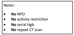
Have a look at the Regions Hospital solid organ injury protocol using the link below. It has not included serial hemoglobin levels for 18 years, since it was written. Take care to look at the little NO box on the left side of the page.

I’d love to hear from anyone who has also abandoned this little remnant of the past. Unfortunately, I think you are in the minority!

Reference: Serial hemoglobin monitoring in adult patients with blunt solid organ injury: less is more. J Trauma Acute Care Open 5:3000446, 2020.

Serial Hemoglobin / Hematocrit – Huh? Part 1

The serial hemoglobin (Hgb) determination. We’ve all done them. Not only trauma professionals, but also other in-hospital clinical services as well. But my considered opinion is that they are not of much use. They inflict pain. They wake patients up at inconvenient hours. And they are difficult to interpret. So why do them?

I’m reposting this mini-series on serial hemoglobin draws in light of a new paper that was published in the Journal of Trauma Nursing. It continues next week with Part 2, and then a summary of the new paper.

First, what’s the purpose? Are you looking for trends or for absolute values? In trauma, the most common reason to order is “to monitor for bleeding from that spleen laceration” or some other organ or fracture complex. But is there some absolute number that should trigger an alarm? If so, what is it? The short answer is, there is no such number. Patients start at a wide range of baseline values, so it’s impossible to know how much blood they’ve lost using an absolute value. And we don’t use a hemoglobin or hematocrit as a failure criterion for solid organ injury anymore, anyway.

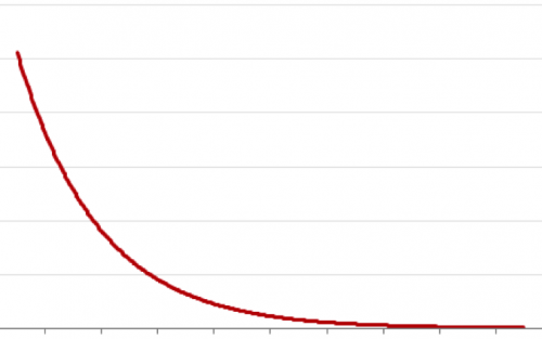
What about trends, then? First, you have to understand the usual equilibration curve of Hgb/Hct after acute blood loss. It’s a hyperbolic curve that reaches equilibrium after about 3 days. So even if your patient bled significantly and stopped immediately, their Hgb will drop for the next 72 hours anyway. If you really want to confuse yourself, give a few liters of crystalloid on top of it all. The equilibration curve will become completely uninterpretable!

And how often should these labs be drawn? Every 6 hours (common)? Every 4 hours (still common)? Every 2 hours (extreme)? Draw them frequently enough, and you can guarantee eventual anemia.

Bottom line: Serial hemoglobin/hematocrit determinations are nearly worthless. They cost a lot of money, they disrupt needed rest, and no one really knows what they mean. For that reason, my center does not even include them in our solid organ injury protocol. If bleeding is ongoing and significant, we will first look at vital signs and a good old physical exam. But if you must, be sure to explicitly state what you will do differently at a certain value or trend line. If you can’t do this and stick to it, then you shouldn’t be ordering these tests in the first place!

In my next post, I’ll discuss a paper that objectively shows the (lack of) utility of this testing method.

Crafting And Refining Your Massive Transfusion Protocol – Part 3

Let’s continue with my series on the massive transfusion protocol (MTP). I’ll continue today with information on deactivating and analyzing your MTP.

Deactivation. There are two components to this: recognizing that high volume blood products are no longer needed, and communicating this with the blood bank. As bleeding comes under surgical control, and CBC and clotting parameters (and maybe TEG/ROTEM) normalize, the pace of transfusion slows, and ultimately stops. Until this happens, the MTP must stay active. Even a low level of product need should be met with coolers stocked with the appropriate ratios of products.

There are two ways to stop the MTP: the surgeon or their surrogate calls the blood bank (when no more blood products are to be used), or the blood bank calls the surgeon after the next cooler has been waiting for pickup for a finite period of time. This is typically about 30 minutes. It is extremely helpful if the exact deactivation time is recorded in the electronic medical record. However, this information can be obtained from the blood bank.

Analysis. It’s all over, and now the real fun begins. For most trauma centers, the blood bank maintains extensive data about every aspect of each MTP event. They record what units were released and when, when they were returned, which ones were used, were they at a safe temperature on return or were they wasted, and much, much more! Typically, one of the blood bank supervisors or a pathologist then compiles and reviews this data. What happens next varies by hospital.

Ideally, the information from every MTP activation gets passed on to the trauma program. Presentation at your transfusion committee is fine, but this data is most suitable for presentation at the trauma operations committee. And if significant variances are present (e.g. product ratios are way off) then it should also be discussed at your multidisciplinary trauma PI committee as well.

There are relatively few standard tools out there that allow the display of MTP data in an easily digestible form. Here are some of the key points that must be reviewed by the trauma PI program:

  • Demographics
  • Components used (for ratio analysis)
  • Lab values (INR, TEG, Hgb, etc)
  • Logistics
  • Waste

I am aware of two tools, the Broxton form and an MTP audit tool from the Australian National Blood Authority. The Broxton tool covers all the basics and includes some additional data points that cover activation criteria, TXA administration, and administration of uncrossmatched blood. Click here to check it out. The Australian tool is much more robust with more data points that make a lot of sense. You can download a copy by clicking here.

In the next post, I’ll continue with activation criteria for the MTP.

Crafting And Refining Your Massive Transfusion Protocol – Part 2

My series on the massive transfusion protocol (MTP) continues! Today, I’ll provide some tips on the logistics of your MTP.

MTP logistics include details such as who will be delivering the blood, what actually goes in each cooler, what ratios should be used, limitations imposed by the use of frozen plasma, and documentation. I’ll discuss details about ratios and FFP in the next Trauma MedEd newsletter.

The runners who travel between the blood bank and the patient need to be selected carefully. Blood bank tech? Not ideal because they’ve got more important work to do. ED or OR tech? Maybe, as long as you’ve got a reliable pool. Student or resident? Probably not, because they may not know their way to the blood bank, which is typically placed in the farthest corner of the basement as possible. One of the most creative solutions I’ve seen is the use of a hospital security officer. Think about it. They know the hospital layout cold, including that obscure corner where the blood bank is located. And there are plenty around all the time!

Documentation is critically important, both in the trauma bay and the blood bank. Trauma activations, especially ones requiring MTP, are very fast-moving and complex. Two sets of documentation are crucial: accurate records of blood product administration (on the trauma flow sheet), and documentation of just about everything else (in the blood bank). A specific timestamp on the trauma flow sheet that records the exact time of activation of MTP is a big plus.

What about coolers? I’ve seen everything used from uninsulated plastic buckets, picnic coolers, and pneumatic tube containers to large, self-contained rolling refrigeration units. The choice of container really boils down to cost vs waste. The cheaper the container is, the less insulated it is, the more likely that blood products will be discarded due to high temperature if not transfused promptly. The best blend of cost vs utility seems to be the good, old-fashioned picnic cooler. It’s very portable, reasonably cheap, and can be tested for temperature maintenance. Just be sure to secure a pouch to the outside to keep platelets at room temperature to maintain full functionality.

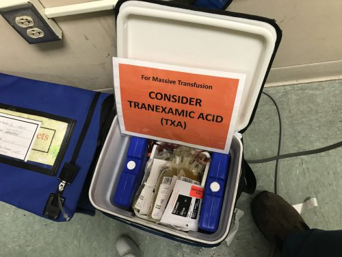
Here’s a sample MTP cooler that’s ready for use. The platelets are in the pouch on the left. Note the reminder to prompt the team to give TXA if not contraindicated. This is often forgotten in the heat of the trauma activation.

Who actually runs your MTP? In some ways, it should run itself. Coolers get delivered, products get transfused. However, some decision making is needed to decide how long to continue and whether any tweaking of product ratios needs to happen. In the emergency department, the surgeon or emergency physician can do this. But once a trauma patient arrives in the OR, that is no longer the case. The emergency physician was left behind in the ED and the surgeon is up to her elbows in trouble. What about the anesthesiologist? Nope, they are busy keeping the patient safely asleep, regulating rapid infusions, and actually administering the blood.

Most of the time, however, these two physicians actually end up running the MTP while multi-tasking at their other job. Unfortunately, this can lead to errors and delays. One best practice to consider: a highly trained trauma resuscitation nurse or advanced practice provider (APP) can run the MTP from the sidelines. They travel with the patient from ED to OR, managing the MTP the entire time. This offloads responsibility from busier people.

In the next post, I’ll continue with MTP deactivation and analysis.