Video: How To Reduce An Ankle Dislocation

Here’s another fun video. It’s directed to emergency physicians and orthopedic surgeons who have to manage ankle dislocations. It will show you the following:

  • Types of ankle dislocation
  • Reduction
  • Splinting
  • The Quigley maneuver
  • How to apply the Sugartong splint
  • Lots of practical tips!

The video was broadcast at a previous Trauma Education: The Next Generation conference, and features Sarah Anderson MD, an orthopedic surgeon at Regions Hospital.

YouTube player

The Medical Orthopaedic Trauma Service

Our population is aging, and falls continue to be a leading cause of injury and morbidity in the elderly. Unfortunately, many elders have significant medical conditions that make them more likely to suffer unfortunate complications from their injuries and the procedures that repair them.

More and more hospitals around the world are applying a more multidisciplinary approach than the traditional model. One example is the Medical Orthopaedic Trauma Service (MOTS) at New York-Presbyterian Hospital/Weill Cornell Medical Center. Any elderly patient who has suffered a fracture is seen in the ED by both an emergency physician and a hospitalist from the MOTS team. Once in the hospital, the hospitalist and orthopaedic surgeon try to determine the reason for the fall, assess for risk factors such as osteoporosis, provide comprehensive medical management, provide pain control, and of course, fix the fracture.

This medical center published a paper looking at their success with this model. They retrospectively reviewed 306 patients with femur fractures involving the greater trochanter. They looked at complications, length of stay, readmission rate and post-discharge mortality. No change in length of stay was noted, but there were significantly fewer complications, specifically catheter associated urinary tract infections and arrhythmias. The readmission rate was somewhat shorter in the MOTS group, but did not quite achieve significance with regression analysis.

Bottom line: This type of multidisciplinary approach to these fragile patients makes sense. Hospitalists, especially those with geriatric experience, can have a significant impact on the safety and outcomes of these patients. But even beyond this, all trauma professionals need to look for and correct the reasons for the fall, not just fix the bones and send our elders home. This responsibility starts in the field with prehospital providers, and continues with hospital through the entire inpatient stay.

Related post:

Reference: The medical orthopaedic service (MOTS): an innovative multidisciplinary team model that decreases in-hospital complications in patients with hip fractures. J Orthopaedic Trauma 26(6):379-383, 2012.

How To Remember Those “Classes of Hemorrhage”

The Advanced Trauma Life Support course lists “classes of hemorrhage”, and various other sources list a similar classification for shock. I’ve not been able to pinpoint where these concepts came from, exactly. But I am sure of one thing: you will be tested on it at some point in your lifetime.

Here’s the table used by the ATLS course:

classes_of_shock

The question you will always be asked is:

What class of hemorrhage (or what % of blood volume loss) is the first to demonstrate systolic hypotension?

This is important because prehospital providers and those in the ED typically rely on systolic blood pressure to figure out if their patient is in trouble.

The answer is Class III, or 30-40%. But how do you remember the damn percentages?

multiscore-maxi1

It’s easy! The numbers are all tennis scores. Here’s how to remember them:

Class I up to 15% Love – 15
Class II 15-30% 15 – 30
Class III 30-40 30 – 40
Class IV >40% Game (almost) over!

Bottom line: Never miss that question again!

Can Prehospital Providers Accurately Estimate Blood Loss? Part 2

I’ve previously written about the difficulties estimating how much blood is on the ground at the trauma scene. In general, EMS providers underestimated blood loss 87% of the time. The experience level of the medic was of no help, and the accuracy actually got worse with larger amounts of blood lost!

A group in Hong Kong developed a color coded chart (nomogram) to assist with estimation of blood loss at the scene. It translated the area of blood on a non-absorbent surface to the volume lost. A convenience study was designed to judge the accuracy that  could be achieved using the nomogram. Sixty one providers were selected, and estimated the size of four pools of blood, both before and after a 2 minute training session on the nomogram.

Here’s what it looks like:

Note the areas across the bottom. In addition to colored square areas, the orange block is a quick estimate of the size of a piece of paper (A4 size since they’re in Hong Kong!)

Here are the factoids:

  • The 61 subjects had an average of 3 years of experience
  • Four scenarios were presented to each: 180ml, 470ml, 940ml, and 1550ml. These did not correspond exactly to any of the color blocks.
  • Before nomogram use, underestimation of blood loss increased as the pool of blood was larger, similar to the previous study
  • There was a significant increase in accuracy for all 4 scenarios using the nomogram, and underestimation was significantly better for all but the 940ml group
  • Median percentage of error was 43% before nomogram training, vs only 23% after. This was highly significant.

Bottom line: This is a really cool idea, and can make estimation of field blood loss more accurate. All the medic needs to do is know the length of their shoe and the width of their hand in cm. They can then estimate the length and width of the pool of blood and refer to the chart . Extrapolation between colors is very simple, just look at the line. The only drawback I can see occurs when the blood is on an irregular or more absorbent surface (grass, inside of a car). 

Related posts:

Reference:  Improvement of blood loss volume estimation by paramedics using a pictorial nomogram: a developmental study. Injury article in press Oct 2017.

What Is: Bucket Handle Injury Of The Intestine

A bucket handle injury is a type of mesenteric injury of the intestine. The intestine itself separates from the mesentery, leaving a devascularized segment of bowel that looks like the handle on a bucket (get it?).

These injuries can occur after blunt trauma to the abdomen. The force required is rather extreme, so the usual mechanism is motor vehicle crash. In theory, it could occur after a fall from a significant height, and I have seen one case from a wood fragment that was hurled at the abdomen by a malfunctioning lathe.

The mechanics of this injury are related to fixed vs mobile structures in the abdomen. Injuries tend to occur adjacent to areas of the intestine that are fixed, such as the cecum, ligament of Treitz, colonic flexures and rectum. During sudden deceleration, portions of the intestine adjacent to these areas continue to move, pulling on the nearby attachments. This causes the intestine itself to pull off of its mesentery.

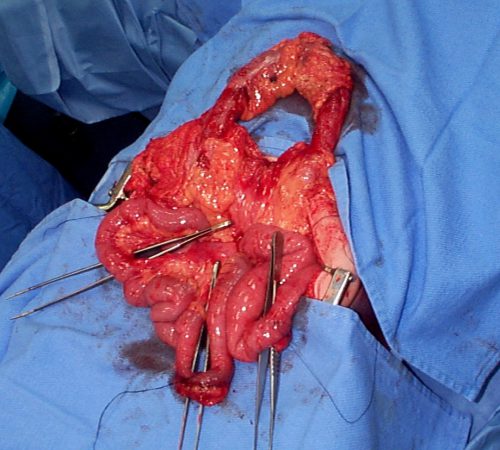
Source: personal archive. Not treated at Regions Hospital

The picture above shows multiple bucket handle injuries in one patient. There are 3 injuries in the small bowel, and one involving the entire transverse colon. Note the obviously devascularized segment at the bottom center of the photo.

The terminal ileum is the most common site for bucket handle tears. Proximal jejunum, transverse colon, and sigmoid colon are other possible areas. In my experience, the driver is more likely to sustain an injury to the terminal ileum, and the front passenger to the sigmoid. I think this is due to the location of the buckle, but it’s just my own observation.

Always think about the possibility of this injury in patients with very high speed decelerationSeat belt marks have a particularly high association with this injury. If your patient has an abnormal exam in the right lower quadrant, or if the CT shows unusual changes there (“dirty” mesenteric fat, thickened bowel wall, extravasation), I recommend a trip to the OR. In these cases, an injury will nearly always be present. And since the affected intestine may take a few days to die and leak, look for an unexplained rise in WBC beginning on hospital day 2.

Related posts: