Tag Archives: shock

ACS Trauma Abstract #6: Scanning Unstable Trauma Patients???

If you’ve read my stuff for very long, you know I frown on sending unstable patients anywhere but to the OR. Instability tends to get worse, and that always happens at inopportune locations like hallways, elevators, and CT scanners. Imagine my surprise when I noticed an abstract being presented at the Clinical Congress of the American College of Surgeons this week suggesting that it was okay to scan hemodynamically unstable patients before “definitive therapy.”

Here’s the title:

“Computed tomography in hemodynamically abnormal thoracoabdominal trauma safely enhances surgical triage”

The devil is in the details and the language. This group from USC included all patients who were hemodynamically abnormal on arrival to the trauma bay but who normalized to SBP > 90 during the resuscitation were included.  A total of 253 of these patients were reviewed over a 9 year period, and the usual variables were analyzed (mortality, complications, hospital, ICU, and vent days, etc).

Here are the factoids:

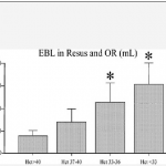
  • Of the 253 patients studied, 45 went to straight to OR and 208 were taken to CT
  • Injury severity was identical for the two groups
  • Lengths of stay and mortality were not different, but only p values were given
  • Patients taken to CT cleared their lactic acidosis faster (12 vs 5 hours), and used a bit less plasma and significantly less blood transfusions
  • The OR group underwent more procedures (31% vs 13%), although what these were and when they were performed is not listed

Bottom line: The title of this abstract is misleading, and may fool those who don’t read the rest of the abstract. It should read:

“Computed tomography in previously hemodynamically abnormal thoracoabdominal trauma safely enhances surgical triage”

Someone who just skims through this issue of the journal may get the idea that it’s okay to scan an unstable patient. The authors are not saying this at all. If you read the conclusion carefully, you can see that the patients had to be resuscitated to a SBP > 90 before they considered taking to scan. And they did that for the majority of these patients.

The real question is, why do the scanned patients clear their lactic acidosis faster, need less blood, and undergo fewer procedures? It appears that there is some bias or selection process in play. Otherwise, why not use the magic CT scanner to make them all better?

Reference: Computed tomography in hemodynamically abnormal thoracoabdominal trauma safely enhances surgical triage. JACS 225(4S2):e175-176, 2017.

EAST 2016: (F)utility of CPR In Hemorrhagic Shock

Ahh, another (f)utility study. Does it work, or doesn’t it? And yes, I know. It’s another animal study. But it may give us a glimpse of where we are really going with this. 

A team at the University of Tennessee – Knoxville devised a dog experiment to study how well performing CPR works in critically hypovolemic animals. They used three groups of dogs that received a severe shock insult: hemorrhage until loss of pulse, then waiting for 30 minutes in that pulseless state. At that point, one of three interventions was performed for 20 minutes.

One group received CPR only, another group underwent CPR plus fluid administration, and the last group got fluids only

Here are the factoids:

  • The insult to all three groups was similar.
  • Vital signs and lab studies were similar in the CPR+fluid and fluid only groups.
  • The CPR only group had significantly lower mean arterial pressures and higher pulse rates than the other CPR+fluid and fluid only groups.
  • Ejection fraction was lower in the CPR only group, and it also had a higher incidence of end organ damage. 
  • Two of the six dogs in the CPR only group died before the end of the study.

Bottom line: Tread with caution here. It makes sense that pounding on an empty tank won’t do much. But this study doesn’t exactly prove this. Only the vital signs measurements were significantly different. All other results are just trends in this very small study. And finally, dogs are (obviously) different than people, in their physiology and their chest wall shape. This can certainly make a difference, and does not mean that we should abandon CPR in humans in hemorrhagic shock.

Reference: Utility of CPR in hemorrhagic shock, a dog model. EAST 2016 Oral abstract #8, resident research competition.

Does Initial Hematocrit Predict Shock?

Everything you know is WRONG!

The classic textbook teaching is that trauma patients bleed whole blood. And that if you measure the hematocrit (or hemoglobin) on arrival, it will approximate their baseline value because not enough time has passed for equilibration and hemodilution. As I’ve said before, you’ve got to be willing to question dogma!

The trauma group at Ryder in Miami took a good look at this assumption. They drew initial labs on all patients requiring emergency surgery within 4 hours of presentation to the trauma center. They also estimated blood loss in the resuscitation room and OR and compared it to the initial hematocrit. They also compared the hematocrit to the amount of crystalloid and blood transfused in those areas.

Patients with lower initial hematocrits had significantly higher blood loss and fluid and blood replacement during the initial treatment period. Some of this effect may be due to the fact that blood loss was underestimated, or that prehospital IV fluids diluted the patient’s blood counts. However, this study appears sound and should prompt us to question the “facts” we hear every day.

Bottom line: Starling was right! Fluid shifts occur rapidly, and initial hematocrit or hemoglobin may very well reflect the volume status of patients who are bleeding rapidly. If the blood counts you obtain in the resuscitation room come back low, believe it! You must presume your patient is bleeding to death until proven otherwise.

Reference: Initial hematocrit in trauma: A paradigm shift? J Trauma 72(1):54-60, 2012.

Clinical Tip: The Flat Vena Cava in Blunt Trauma

Trauma patients who are hypotensive in the Emergency Department can only be transported to one of two places: the operating room or the morgue. With rare exception, they should never be taken outside the department (e.g. CT scan) because of the fear that they may arrest in an area that is not conducive to efficient resuscitation.

Sometimes patients are initially stable but decompensate later. Since most stable blunt trauma patients end up in CT scan, perhaps there is some telltale sign that can predict later deterioration. A recent Japanese paper looked at the “flatness” of the inferior vena cava as seen on the abdominal CT scan as a predictor of hemodynamic decompensation in the first 24 hours.

A small cohort of 114 patients was used in this prospective study. The vena cava was evaluated at the level of the renal veins. The flatness of the IVC was determined by dividing the transverse diameter by the anteroposterior (AP) diameter. A flat IVC was defined as a transverse to AP diameter ratio of more than 4:1. The ratio in normal patients was about 2:1. See the figure for details.

Patients who had a flat IVC required significantly more blood transfusions, crystalloid infusions within 2 hours of admission, and were more likely to proceed to the OR within the first 24 hours of their hospital stay.

Bottom Line: Assuming that you are only taking stable blunt trauma patients to CT, the incidental finding of a flat vena cava should increase your paranoia levels and lower your threshold for ordering blood and getting the trauma surgeons involved. 

Reference: Predictive value of a flat inferior vena cava on initial computed tomography for hemodynamic deteroration in patients with blunt torso trauma. J Trauma 69(6):1398-1402, 2010.