Tag Archives: hemothorax

Retained Hemothorax Part 2: Lytics (again)

Yesterday, I reviewed a small case report that was published a couple of years ago on lytics for treatment of retained hemothorax. But surely, there must be something better, right?

After digging around, I did find a paper from 2007 that prospectively looked at protocolized management of retained hemothorax, and its aftermath. It was carried out at a busy Level I trauma center over a 16 month period.

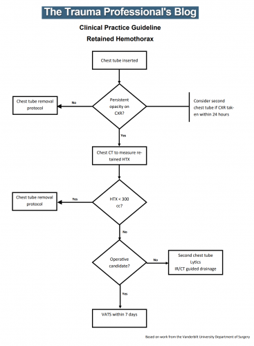
All patients with a hemothorax treated with chest tube received daily chest x-rays. Those with significant opacification on day 3 underwent CT scan of the chest. If more than 300 cc of retained blood was present, the patient received streptokinase or urokinase (surgeon preference and drug availability) daily, and rolled around in bed for 4 hours to attempt to distribute it. The chest tube was then unclamped and allowed to drain. This was repeated for 3 days, and if there was still opacification, a repeat CT was obtained. If the volume was still greater than 300 cc, the cycle was repeated for the next 3 days. If the opacification cleared at any point, or the repeat CT showed less than 300 cc, the protocol was stopped and the chest tube removed. If the chest was still opacified after 6 days, VATS was offered.

Here are the factoids:

  • A total of 203 patients with hemothorax were admitted during the study period and 25 (12%) developed a retained hemothorax
  • While a few had treatment start within 4 days, the majority did not receive lytics until day 9 (range 3  –30 days!)
  • The average length of time in hospital after start of lytics was 7 days, leading to a total length of stay of 18 days
  • 92% of patients had “effective” evacuation of their retained hemothorax, although 1 had VATS anyway which found only 100 cc of fluid
  • 16 patients had “complete” evacuation, and 5 had “partial” evacuation
  • There were no hemorrhagic complications, but one third of patients reported significant pain with drug administration

Bottom line: Sounds good, right? The drug seems reasonably effective, although lengths of stay are relatively long. However, streptokinase and urokinase are no longer available in the US, having been replaced with tissue plasminogen activator (tPA). This paper does a cost analysis of lytics vs VATS and found that the former treatment cost about $15000 (drug + hospital stay) vs $34000 for VATS. However, a big part of this was that the drug only cost about $75 per dose. tPA is much more expensive.

So once again, small series, longer lengths of stay, but at least nicely done. Unfortunately, the drug choice is no longer available so use of tPA tilts the balance away from lytics. 

Reference: Intrapleural Thrombolysis for the Management of Undrained Traumatic Hemothorax: A Prospective Observational Study. J Trauma 62(5):1175-1179, 2007.

Retained Hemothorax Part 1: Lytics

Hemothorax is a common complication of chest trauma, occurring in about one third of cases. It is commonly treated with a chest tube, which usually takes care of the problem. But in a few cases some blood remains, which can result in an entrapped lung or empyema.

There are several management options. Historically, these patients underwent thoracotomy to peel out the fibrinous collection stuck to lung and chest wall. This has given way to the more humane VATS procedure (video assisted thoracoscopic surgery) which accomplishes the same thing using a scope. In some cases, another tube can be inserted, sometimes under CT guidance, to try to drain the blood.

So what about lytics? It’s fibrin, right? So why not just dissolve it with tissue plasminogen activator (tPA)? There have been very few studies published over the years. The most recent was in 2014. I’ll review it today, and another tomorrow. Finally, I’ll give you my thoughts on the best way to deal with retained hemothorax.

Here are the factoids:

  • This was a single center, retrospective review of data from 1.5 years beginning in 2009
  • A total of seven patients were identified, and most had hemothorax due to rib fractures. Three presented immediately after their injury, 4 were delayed.
  • Median time from injury to chest tube placement was 11 days
  • Median time the chest tube was in place was 13 days, with an average hospital stay of 14 days
  • Patients received 1 to 5 treatments, averaging 24mg per dose
  • There was one death in the group, unrelated to TPA treatment
  • No patient “required” VATS, but one underwent thoracotomy, which turned out to be for a malignancy

Bottom line: The authors conclude that tPA use for busting retained hemothorax is both safe and effective. Really? With only seven patients? The biggest problem with this study is that it uses old, retrospective data. We have no idea why these patients were selected for tPA in this 5-year old cohort of patients. Why did it take so long to put in chest tubes? Why did the chest tubes stay in so long? Maybe this is why they were in the hospital so long?

Plus, tPA is expensive. A 100mg vial runs about $6000. Does repeatedly using an expensive drug and keeping a patient in the hospital an extra week or so make financial sense? So it better work damn well, and this small series doesn’t demonstrate that.

Tomorrow, I’ll look at the next most recent paper on the topic, from way back in 2004.

Reference: Evaluation of chest tube administration of tissue plasminogen activator to treat retained hemothorax. Am J Surg 267(6):960-963, 2014.

EAST 2019 #4: Predicting Retained Hemothorax

Traumatic hemothorax is an interesting problem. Sometimes it clots, sometimes it doesn’t. Sometimes it sticks to the lung and causes a restrictive problem, sometimes it doesn’t. And even if it doesn’t fuse to the lung, having a moderate volume of retained blood can create significant functional problems.

What if we could predict which hemothoraces would progress to retained hemothorax so we could intervene sooner and avoid complications and potential wasted time in the hospital? An EAST multi-institutional trial explored a possible technique for doing just that, postulating that an initial large volume hemothorax (HTX) would correlate with development of retained HTX. This study used data collected prospectively from 17 US trauma centers, and involved the use of Mergo’s formula to calculate the size of the HTX. This technique essentially estimates the volume of a cylinder that fits inside the largest diameter of the HTX seen on CT scan, extending the entire length of the thorax. Although this technique will underestimate the volume at the largest part of the HTX, it overestimates it above and below this point, so it probably all evens out. Roughly.

Here are the factoids:

  • 985 patients were enrolled and 1033 HTX were measured
  • Patients who developed retained HTX (defined as blood in the chest requiring intervention or subsequent identification of HTX by CT) were more likely to have penetrating trauma, pulmonary contusion, and had their HTX seen on initial chest x-ray (CXR)
  • Chest abbreviated injury score (AIS) was higher in patients with retained HTX, and their calculated HTX volume was larger
  • Chest tube size did not correlate with development of retained HTX
  • The authors concluded that patients who developed retained HTX had a lower initial hematocrit, higher chest AIS, and large volume seen on CT

Here are some questions for the authors and presenter to consider in advance to help them prepare for audience questions:

  • When and over what period of time was this data collected? It was not stated in the abstract. Very old or long-running data sets run the risk of technology (CT scan resolution) or practice (pigtail vs chest tube) changes.
  • Couldn’t the hematocrit and chest AIS factors be red herrings? Did this study look at all the other injuries that could have decreased the hematocrit or just focus on the chest injury? And the chest AIS automatically increases from 3 to 4 as the volume of the HTX increases. Could this skew your analysis?
  • One of the definitions of retained HTX (need for intervention) depends on clinical judgement and practice habits, which probably differ at the various trauma centers enrolled.
  • Only odds ratios were listed in the abstract. Even though the HCT and AIS numbers are statistically significantly different, I can’t tell if they are clinically significant. Be sure to share your means and standard deviation/error during your presentation.
  • Did you lump pigtail catheters in with regular chest tubes? And did you look at the distribution of chest tube sizes? If all were large it would be hard to come to a good conclusion about size.
  • What about trauma centers who do not use CT on every trauma patient? Do we really need to CT someone who falls down, breaks a few ribs, and has a visible HTX on CXR?
  • Big picture: so what do we do now based on your findings? How do we plan re-imaging and potential surgical intervention in these patients?

This is very interesting work, and I look forward to the presentation!

Reference: Predictors of post-traumatic retained hemothorax: results of an EAST multi-institutional trial. EAST 2019 Paper #14.

Chest Tubes: Size Doesn’t Matter – Part 2

A few days ago, I wrote about a paper that seemed to suggest that using a smaller chest tube (28-32 Fr) vs larger ones (36-40 Fr). The results suggested that their function was very similar. I emphasized that I thought the result was intriguing, because I’m of the opinion that bigger is better for getting clotted blood out. However, I am amenable to changing my mind based on newer, better data.

But I did caution readers that I would like to see more data. One study should never change your practice! Then I see a lot of chatter on Twitter about another study from 2016 that looks at even smaller tubes, with people saying they will now switch to pigtail catheters (12 Fr)!!

First, not a logical progression of thinking there. And second, let’s take an actual look at the paper. It’s from an emergency medicine group in Fukui, Japan, which retrospectively reviewed their 7 year experience with using a small (20-22 Fr) vs large (28 Fr) tubes. They identified a total of 124 chest tube insertions to compare, 68 small and 56 large.

Now let’s look at the factoids:

  • Demographics, mechanism, and ISS were the same between groups
  • Duration of insertion and initial drainage were also the same between groups
  • Complication rates were similar, with 1 empyema and 2 retained hemothoraces in each group
  • Additional tubes were place in 2 patients with small tubes vs 4 with large tubes
  • Thoracotomy was performed in 2 patients with small tubes vs 1 with a large tube

Based on all of this, the authors concluded that there was no difference in drainage efficacy, complications, or need for additional invasive procedures.

Wait a minute!! Again, if you only read the abstract, you might be led to start using ever smaller chest tubes. But read the entire paper! There are many problems with this paper, including:

  • It’s a very small, retrospective review. This automatically means that the statistical power is suspect.
  • Why did they only document 124 insertions over 7 years?? That’s about one every 3 weeks! Either a lot of data are missing or they are not very busy. But Fukui Prefectural Hospital has over 1000 beds! So it’s the former, not the latter.
  • The retrospective nature means it is not possible to determine why a particular tube size was chosen. Roll of the dice? This fact alone introduces a huge potential for selection bias. Was a smaller tube selected because the hemothorax looked smaller? Probably! The fact that 4 patients with larger tubes had another one placed suggests that they were being used for larger collections. And patients with higher ISS tended to get bigger tubes.

Bottom line: Don’t change your practice based on this paper. And certainly don’t choose to use even smaller pigtails. And of course, always critically read any paper that you like to make sure you are not cherry picking the ones you choose to believe. IMHO, it’s still best to use big (36 Fr) or bigger (40 Fr).

Reference: Small tube thoracostomy (20-22 Fr) in emergent management of chest trauma. Injury 48:1884-1887, 2016.

Retained Hemothorax Part 4: The Practice Guideline

Over the last three days, I reviewed some data on lytics at the request of some of my readers. Then I looked at a paper describing one institution’s experience dealing with retained hemothorax, including the use of VATS. But there really isn’t much out there on how to roll all this together.

Until now. The trauma group at Vanderbilt has a paper in press describing their experience with a home-grown practice guideline for managing retained hemothorax.  Here’s what it looks like:

I know it’s small, so just click it to download a pdf copy. I’ve simplified the flow a little as well.

All stable patients with hemothorax admitted to the trauma service were included over a 2.5 year period. The practice guideline was implemented midway through this study period. Before implementation, patients were treated at the discretion of the surgeon. Afterwards, the practice guideline was followed.

Here are the factoids:

  • There were an equal number of patients pre- and post-guideline implementation (326 vs 316)
  • An equal proportion of each group required an initial intervention, generally a chest tube (69% vs 65%)
  • The number of patients requiring an additional intervention (chest tube, VATS, lytics, etc) decreased significantly from 15% to 9%
  • Empyema rate was unchanged at 2.5%
  • Use of VATS decreased significantly from 8% to 3%
  • Use of catheter guided drainage increased significantly from 0.6% to 3%
  • Hospital length of stay was the same, ranging from 4 to 11 days (much shorter than the lytics studies!)

Bottom line: This is how design of practice guidelines is supposed to work. Identify a problem, typically a clinical issue with a large amount of provider care variability. Look at the literature. In general, find it of little help. Design a practical guideline that covers the major issues. Implement, monitor, and analyze. Tweak as necessary based on lessons learned. If you wait for the definitive study to guide you, you’ll be waiting for a long time.

This study did not significantly change outcomes like hospital stay or complications. But it did decrease the number of more invasive procedures and decreased variability of care, with the attendant benefits from both of these. It also dictates more selective (and intelligent) use of additional tubes, catheters, and lytics. 

I like this so much that I plan to adopt it at my center!

Download the practice guideline here.

Related posts:

Posts in this series:

Reference: Use of an evidence-based algorithm for patients with traumatic hemothorax reduces need for additional interventions. J Trauma, in press, December 14, 2016.