Tag Archives: hemothorax

Retained Hemothorax Part 3: VATS

I’ve written about the use of lytics to treat retained hemothorax over the past few days. Although it sounds like a good idea, we just don’t know that it works very well. And they certainly don’t work fast. Lengths of stay were on the order of two weeks in both studies reviewed.

The alternative is video assisted thoracoscopic surgery (VATS). So let’s take a look at what we know about it. This procedure is basically laparoscopy of the chest. A camera is inserted, and other ports are added to allow insertion of instruments to suck, peel, and scrape out the hemothorax.
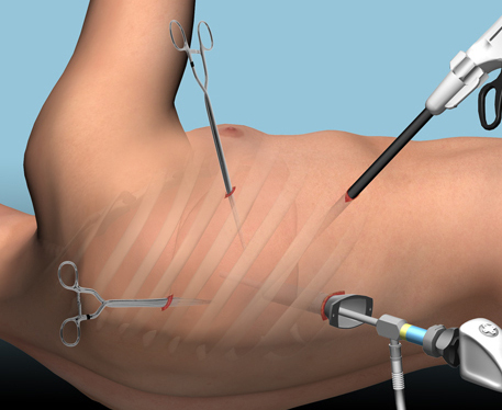
A prospective, multi-center study was performed over a 2 year period starting in 2009. Twenty centers participated, contributing data on 328 patients with retained hemothorax. This was defined as CT confirmation of retained blood and clot after chest tube placement, with evidence of pleural thickening.

Here are the factoids:

  • 41% of patients had antibiotics given for chest tube placement (this is interesting given the lack of consensus regarding their effectiveness!)
  • A third of patients were initially managed with observation, and most of them (82%) did not need any further procedures (83 of 101 patients)
  • Observation was more successful in patients who were older, had smaller hemothoraces (<300cc), smaller chest tubes (!!, <34 Fr), blunt trauma, and peri-procedure antibiotics (?)
  • An additional chest tube was inserted in 19% of patients, image guided drain placement in 5%, and lytics in 5%. Half to two-thirds of these patients required additional management.
  • VATS was used in 34% of patients. One third of them required additional management including another chest tube, another VATS, or even thoracotomy.
  • Thoracotomy was most likely required if there was a diaphragm injury or large hemothorax (<900cc)
  • Empyema and pneumonia were common (27% and 20%, respectively)

Bottom line: There’s a lot of data in this paper. Most notably, many patients resolve their hemothorax without any additional management. But if they don’t, additional tubes, guided drain placement, and lytics work only a third of the time and contribute to additional time in the hospital. Even VATS and thoracotomy require additional maneuvers 20-30% of the time. And infectious complications are common. This is a tough problem!

Tomorrow, I’ll try to roll it all together and suggest an algorithm to try to optimize both outcomes and cost.

Posts in this series:

Reference: Management of post-traumatic retained hemothorax: A prospective, observational, multicenter AAST study. 72(1):11-24, 2012.

Retained Hemothorax Part 1: Lytics

Hemothorax is a common complication of chest trauma, occurring in about one third of cases. It is commonly treated with a chest tube, which usually takes care of the problem. But in a few cases some blood remains, which can result in an entrapped lung or empyema.

There are several management options. Historically, these patients underwent thoracotomy to peel out the fibrinous collection stuck to lung and chest wall. This has given way to the more humane VATS procedure (video assisted thoracoscopic surgery) which accomplishes the same thing using a scope. In some cases, another tube can be inserted, sometimes under CT guidance, to try to drain the blood.

So what about lytics? It’s fibrin, right? So why not just dissolve it with tissue plasminogen activator (tPA)? There have been very few studies published over the years. The most recent was in 2014. I’ll review it today, and another tomorrow. Finally, I’ll give you my thoughts on the best way to deal with retained hemothorax.

Here are the factoids:

  • This was a single center, retrospective review of data from 1.5 years beginning in 2009
  • A total of seven patients were identified, and most had hemothorax due to rib fractures. Three presented immediately after their injury, 4 were delayed.
  • Median time from injury to chest tube placement was 11 days
  • Median time the chest tube was in place was 13 days, with an average hospital stay of 14 days
  • Patients received 1 to 5 treatments, averaging 24mg per dose
  • There was one death in the group, unrelated to TPA treatment
  • No patient “required” VATS, but one underwent thoracotomy, which turned out to be for a malignancy

Bottom line: The authors conclude that tPA use for busting retained hemothorax is both safe and effective. Really? With only seven patients? The biggest problem with this study is that it uses old, retrospective data. We have no idea why these patients were selected for tPA in this 5-year old cohort of patients. Why did it take so long to put in chest tubes? Why did the chest tubes stay in so long? Maybe this is why they were in the hospital so long?

Plus, tPA is expensive. A 100mg vial runs about $6000. Does repeatedly using an expensive drug and keeping a patient in the hospital an extra week or so make financial sense? So it better work damn well, and this small series doesn’t demonstrate that.

Tomorrow, I’ll look at the next most recent paper on the topic, from way back in 2004.

Posts in this series:

Reference: Evaluation of chest tube administration of tissue plasminogen activator to treat retained hemothorax. Am J Surg 267(6):960-963, 2014.

Serial Hemoglobin To Monitor Chest Tube Output? Huh?

I’ve already written about the (f)utility of serially monitor hemoglobin (Hgb) or hematocrit (Hct) levels when managing solid organ injury nonoperatively. What about if you are concerned with bloody output from a chest tube drainage system? Could it be of any use there?

Seems like a reasonable idea, right? Wrong. As always, think it through and do the math! Here are the questions you need to ask yourself:

  • What is the Hgb or Hct of the fluid coming out of the chest tube? At worst, it will be the same as the patient, assuming that pure, whole blood is coming out. But this is seldom the case. The fluid is usually described as “serosanguinous”, which is not very exact, but tells you that it is thinner than blood. And if it looks more like Kool-Aid, the concentration is very low indeed.
  • What is the volume in the container? Most collection systems will collect a maximum of 1 to 1.5 L of juice.
  • How fast is it coming out? These things almost never fill right in front of your eyes. It’s a slow process, with less than a few hundred ccs per shift.

Here’s a few hundred ccs of thin drainage in a collection system. Probably decrease in Hgb value – < 0.1, which is far less than the range of lab error.

Bottom line: So now do the math. Let’s say the fluid has half the hematocrit / hemoglobin of whole blood. Losing one unit (500cc) of whole blood will generally drop your Hgb by about 1 gm, or your Hct by about 3%. If the blood is half-strength like I am proposing (and the usual drainage is typically much thinner), it will take twice as much (one liter) loss to drop the lab values by that much. This will probably come close to filling up the average collection system. If it takes a day or two or more to fill up, you are not going to see much change in their lab values. And most of the time, the blood in the system is thin like Kool-Aid, so your patient is really losing very little actual blood.

So measuring serial hemoglobin / hematocrit as you watch a hemothorax drain doesn’t make sense. Unless the output is pure blood and the system is filling up in front of your eyes, of course. In that case, a trip to the OR to fix the problem might be a better idea than doing a blood draw and sitting around waiting for the result to come back.

Related post:

Retained Hemothorax And Empyema

Patients with chest trauma sustain hemothorax on occasion. The trauma professional usually picks this diagnosis up in the initial evaluation and makes a decision whether or not to drain it. The parameters for this decision are not very clear, even today. But what happens when there is residual hemothorax? Should we be more aggressive in getting it out?

All this boils down to an understanding of the natural history of retained hemothorax. This kind of information can help us decide whether to be more aggressive in our efforts to remove it. The results of a multicenter study looking at this issue was published recently. They focused on patients who had a chest tube placed for management of either hemo- or pneumothorax within 24 hours of admission. Patients who had suspected retained hemothorax after tube removal received a CT scan within 14 days. The usual outcomes were studied (length of stay, complications) as well as development of empyema (purulence, acidic pleural fluid, positive Gram stain or culture).

Some interesting results:

  • 328 patients were enrolled across 20 centers. Not a lot, but one of the bigger studies to date.
  • Empyema was diagnosed in 27% of patients
  • Risk factors identified included rib fractures, ISS > 25, and performance of additional interventions for drainage
  • Patients who developed empyema stayed in the ICU and the hospital longer

Bottom line: Retained hemothorax turns into a very serious problem in a quarter of trauma patients who have a chest tube inserted. The presence of residual blood after the chest tube is removed should prompt us to figure out if it’s solid clot or liquid blood (remember the old decubitus view chest xray? They still work!). If it’s liquid, consider drainage via thoracentesis or a smaller catheter. If it’s clot, it may require more invasive techniques to drain it (VATS). If you decide to send the patient home, have them watch out for fevers, chest pain, dyspnea and other symptoms and signs of a developing complication, and make sure they report it to you promptly.

Related post:

Reference: Development of posttraumatic empyema in patients with retained hemothorax: Results of a prospective, observational AAST study. J Trauma 73(3):752-757, 2012.

Extended FAST Exam in Trauma Patients

By now, every emergency medicine physician and surgeon knows what FAST is. This valuable technique allows us to quickly (get it?) determine whether a patient has blood in the abdomen or around the heart which might require operative management. Extended FAST (E-FAST) is an extension of the original technique that allows us to detect the presence of pneumothorax or hemothorax more quickly and accurately than with the conventional chest x-ray.

Both hemothorax and pneumothorax can be missed by x-ray. It takes at least 200cc of free fluid in the chest to show on the chest x-ray, assuming an ideal body habitus. As little as 20cc can be detected using the E-FAST. Studies have also shown that 30-50% of pneumothoraces are missed by x-ray. This diagnostic inaccuracy is due to the fact that hemothoraces settle out posteriorly and pneumothoraces anteriorly. Since the vast majority of chest x-rays in major trauma patients are taken with the patient supine to protect their spine, the bulk of the blood or air have layered out and cannot be seen well. A chest x-ray is still needed, however, to determine injury to the mediastinum and lung parenchyma.

E-FAST exam can be performed by using the standard curvilinear probe. It is usually placed longitudinally on the anterior chest to detect pneumothorax, using the space between two ribs as the “window” to the pleura. The depth setting should be adjusted so that only about 4cm is visible on the display. The junction of the visceral and parietal pleura should be visualized at the backside of the ribs. With a very steady hand, the junction between the two sets of pleura should be scrutinized closely.

If the two sets of pleura slide freely over each other, pneumothorax is unlikely. If not, it may be present. Pneumothorax is not a uniform phenomenon, except when it is of large size. It may be necessary to move the probe to a few other rib spaces to ensure that a smaller pneumothorax is not present.

FALSE POSITIVE ALERT! If the patient is not ventilating well, or if they have a right mainstem intubation, the affected lung(s) may not show the sliding sign, leading the examiner to think they have a problem when they may not.

To detect a hemothorax, the probe is directed upward somewhat when doing the right and left upper abdominal views. A dark triangle located above the diaphragm indicates fluid in the chest (blood). The dark crescent on the left in the image below is a large hemothorax.

E-FAST hemothorax

The bottom line: Extended FAST can be helpful in detecting a significant hemothorax or pneumothorax and can expedite the definitive management of those conditions. If you are already familiar with FAST, a little extra ultrasound training may be very helpful.