Category Archives: Thorax

Detecting Rib Fractures In The Elderly

It’s well known that our elders do less well than younger folks after injury. The number of complications is higher, there tends to be more loss of independence during recovery, and mortality is increased. This is not only true of high energy trauma like car crashes, but also much lower energy events such as a fall from standing.

Rib fractures are common after falls in the elderly and contribute to significant morbidity if not treated adequately. Traditionally, they are identified through a combination of physical exam and chest x-ray. Unfortunately, only half of rib fractures are visible on x-ray. It falls to the physical exam to detect the rest.

A group at Beth Israel Hospital in Boston explored the utility of using chest CT in an attempt to determine if this would result in more appropriate and cost-efficient care in the elderly. They performed a retrospective study of 3 years of their own data on patients aged 65 or more presenting after a mechanical fall and receiving a rib fracture diagnosis. Imaging was ordered at the discretion of the physician. A total of 330 patients were elderly, fell, and had both chest x-ray and chest CT obtained. This was a very elderly group, with a mean age of 84 years!

Here are the factoids:

  • Rib fractures were seen on chest x-ray in 40 patients (12%) and on CT in an additional 56 ; 234 patients had no fractures on either
  • When fractures were seen on both studies, CT identified a median of 2 more fractures than chest x-ray
  • Patients with fractures not seen on chest x-ray were admitted significantly more often than those without fractures (91% vs 78%)
  • Mortality, admission to ICU, ICU length of stay, and hospital length of stay were not different if fractures were seen only on CT
  • CT scan identified new issues or clarified diagnoses suggested by chest x-ray in 14 cases, including one malignancy
  • Rib detail images were obtained in 13 patients and proved to be better than chest x-ray, but not quite as good as CT scan

Conclusion: use of CT for rib fracture diagnosis resulted in a few more admissions, but no change in hospital resource utilization, complications, or mortality.

Bottom line: Hmm…, read the paper closely. The authors conclude that more patients with CT-only identified rib fractures are admitted. But compared to what? Unfortunately, patients without rib fractures on CT. What about comparing to patients who had fractures seen on chest x-ray too? If that number is the same, then of what additional use is CT? Identifying a few incidentalomas?

Given that there is no change in the usual outcome measures listed here, it doesn’t seem like there is any additional benefit to adding CT. And I can see a lot of downsides: cost, radiation, and possible exposure to IV contrast. In my mind, there is still nothing that beats a good physical exam and a chest x-ray. Skip the CT scan. And don’t even think about ordering rib detail images! That’s so 1990s. And even if no rib fractures are seen on imaging, physical exam is the prime determinant for admitting your patient for aggressive pain management and pulmonary toilet.

Reference: Chest CT imaging utility for radiographically occult rib fractures in elderly fall-injured patients. J Trauma ePub ahead of print, Jan 23, 2019.

Print Friendly, PDF & Email

Best of AAST #6: Antibiotics For Chest Tubes??

For as long as I can remember (nearly 50 years worth of literature) there has been some debate about giving antibiotics after chest tube insertion to decrease the infection rate. The pendulum moved back and forth for decades, never getting very far into the “give antibiotics” side. It’s been quite a while since I remember any new papers on this, and I thought the debate had been resolved in favor of never using them.

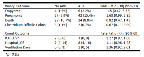
But then I see an abstract from the AAST multi-institutional trials group studying presumptive antibiotics after chest tube insertion! They conducted a prospective, observational study at 22 Level I trauma centers, enrolling nearly 2,000 patients. They matched patients in antibiotic and no antibiotic groups, arriving at (only) 272 patients in each group.

Here are the results:

Bottom line: First, it’s a little disappointing that the numbers were so low with a trial that includes 22 trauma centers. Did they have a hard time finding centers that would give antibiotics? Or was it just hard to match patients for the variables they were looking at? Regardless, there were no significant differences in infectious complications, and a non-clinically significant difference in ICU stay with antibiotics.

Why won’t this die? If there are so few papers that show an actual benefit from giving antibiotics after chest tube insertion with 50 years of data, then it’s very unlikely that it will ever be shown to be necessary!

Reference: Presumptive antibiotics for tube thoracostomy for traumatic pneumothorax. Session XXII Paper 49, AAST 2018.

Print Friendly, PDF & Email

Best of AAST #5: Pneumothorax – How Big Is Too Big?

Deciding when to place a chest tube can be challenging. Sometimes, it’s obvious: there is a large hemo- or pneumothorax staring you in the face on the chest x-ray. But sometimes, it’s there but “not that big.” The real question is, how big is too big.

That’s a question that’s been very difficult to quantify. The authors of this abstract, from the Medical College of Wisconsin, conducted a six-year retrospective review of every patient with an isolated pneumothorax at their Level I trauma center. Based on their previous research, a 35mm threshold was used to stratify patients into two groups. This measurement was obtained from axial images of a CT scan. Statistical analysis was performed to identify the predictive value in determining whether the patient could be managed without a chest tube.

Here are the factoids:

  • A total of 1767 patients had a pneumothorax during the 6-year period, and about half met inclusion criteria for the study
  • Of the 385 with pneumothorax alone, 92% were managed without a chest tube
  • Of those 353, 95% had a maximum chest wall to lung distance (335)
  • The 35mm measurement was statistically shown to be an independent predictor of successful management without a tube for both blunt and penetrating trauma

Bottom line: Not so fast! Although this looks like a slam dunk abstract, it’s really not. First, many (or most?) pneumothoraces are initially diagnosed using a plain old chest x-ray. A 35mm measurement is meaningless here because there can be significant changes in position of the pneumothorax on the image. Sometimes, the air is located anteriorly with little or no lateral component.  Does this mean we should CT every patient with a known or suspected pneumothorax? I think not.

And the second issue is the subjectivity surrounding the definition of a failure. What criteria were used when the tube was actually placed in this series. If every patient had to become symptomatic first, then I might agree. But I suspect the tubes were placed when followup imaging showed that the air was just “too big.” You can’t statistic away this kind of potential bias from subjectivity.

So what’s the answer? Unfortunately, there still isn’t one. The need for a chest tube must still be based on subjective size on a chest x-ray, physiologic status, and the patient’s ability to tolerate a given amount of lost lung function. It continues to boil down to the  assessments of each trauma professional as to “how big is too big.”

Reference: Observing pneumothoraces: the 35mm rule is safe for both blunt and penetrating chest trauma. Session XVA Paper 28, AAST 2018.

Print Friendly, PDF & Email

How Safe Is ED Thoracotomy?

A few weeks ago, I opened a survey to find out common practices regarding performing emergency thoracotomy (EDT) in the emergency department. This procedure is performed at one time or another in most higher level trauma centers. It’s very invasive and is performed in an area that is not really set up for major operative cases. Furthermore, the atmosphere can be chaotic, and stress levels run high.

How safe is this situation? How does personal safety balance out with saving your patient? There are many, many opportunities for injury during this procedure, with significant exposure to blood and other bodily fluids.

A recently published multi-center study examined the potential for exposure during EDT at 16 US trauma centers over a 1.5 year period (14 Level I, 2 Level II). The study was prospective and observational, and was based on questionnaires filled out by all personnel involved in each procedure. A total of 1360 providers submitted information on 305 EDTs.

Here are the factoids:

  • Mechanism was penetrating in 77% of patients, who were predominantly young and male (91%)
  • 15 patients survived (5%), and 4 had residual neurologic impairment
  • Only 56% of respondents wore full personal protective equipment (PPE)
  • There was a 7% exposure rate per EDT(22 incidents), and 1.6% rate per participant in the case
  • The majority of those exposed were trainees (68%) who were injured by something sharp (scalpel 39%, fracture 28%, needle 17%, scissors 3%)
  • There was a strong correlation with PPE use and no exposure during the procedure
  • Only 92% followed their hospital’s occupational exposure protocol if injured (!!!)

Bottom line: Emergency thoracotomy will always be a dangerous procedure. Things happen quickly, there is little time to properly prepare and sharp, pointy things are everywhere. But according to this paper, the actual exposure rate is low. Factoring in the risk of disease transmission, the risk to an individual provider of contracting HIV is 1 in a million, and for hepatitis C is 3 in 100,000

The most distressing part of this study, to me, was the sense of invulnerability of a few of the participants. How can anyone justify not wearing full PPE during an emergency thoracotomy? I believe this represents a very casual attitude toward wearing PPE in any resuscitation. But this study clearly shows a large decrease in exposure rate when full PPEs are worn. Even more disturbing? The fact that 8% chose not to protect themselves by following their own institution’s occupational exposure protocol. Unforgivable!

The main takeaway messages are: always wear your PPE to a trauma resuscitation because you never know when you’ll need to get invasive (and won’t have time to dress up then), and be careful!!

Reference: Occupational exposure during emergency department thoracotomy: A prospective, multi-institution study. J Trauma 85(1):78-84, 2018.

Print Friendly, PDF & Email

Systemic Air Embolism From Chest Trauma

Systemic air embolism (SAE) is an uncommonly encountered but potentially catastrophic complication that trauma professionals will probably see only once or twice in their career. In a couple of very old papers (30-35 years!), it was said to occur in 4 to 14% of patients with severe lung trauma. The pathognomonic finding is complete cardiovascular collapse shortly after intubation and positive pressure ventilation of a patient with significant chest trauma.

SAE can occur after major blunt or penetrating injury. Normally, pressure in the pulmonary arterial and venous branches of the lungs is higher than that of air in the bronchi. If lung parenchyma is torn by gunshot, stab, or blunt pulmonary laceration, then blood can leak into the smaller airways. Unless a major vessel is torn, the volume lost is not of much consequence. A small amount of hemoptysis may be present.

But if positive pressure is applied to the airways, air may be forced into those vascular structures. If the injury involves a pulmonary vein, the gas bubbles enter the left heart and and then the systemic circulation. Only a cc or two of air in the cerebral circulation or coronary arteries can cause a rapidly fatal condition.

How can a trauma professional suspect that a patient may be susceptible to SAE? Look for evidence of hemoptysis in patients with penetrating chest injury or severe blunt chest trauma. Unfortunately, this occurs in only a few patients and its absence is not helpful.

Always suspect SAE if your patient suffers circulatory collapse or arrest shortly after intubation and positive pressure ventilation. This is especially true in patients who were very stable up until that point. Ultrasound may be used to detect air bubbles in the left heart. Transesophageal echo is even better, but not readily available in the ED.

What can be done if your patient (nearly) arrests after intubation and you suspect SAE? The recommended treatment is single lung ventilation and thoracotomy on the injured side. The injured side is obvious in patients with unilateral penetrating injury, but much more difficult to determine in blunt trauma where either lung may be involved. A quick chest x-ray could be obtained, but may not localize the injured lung.

If the left lung is involved, push the endotracheal tube into the right mainstem bronchus to eliminate the abnormal pressure gradient in the injured lung. If the right lung in injured, a dual lumen endotracheal tube should be inserted for single-lung ventilation. Unfortunately, this requires fiberoptic tools and is not available in the field or most emergency departments.

If single-lung ventilation can be accomplished, this may buy some time to try to resuscitate your patient. A thoracotomy of the injured side can also be carried out to occlude the hilum of the lung in an attempt to stop any further embolism. Initial clamping should be carried out by hand, as using a crushing clamp commits the patient to an emergency pneumonectomy (if they survive beyond this point).

Overall survival is dismal. Old data suggests that more than 80% of blunt trauma victims with SAE die, as well as half of patients with penetrating injury. These numbers are even lower once the patient arrests. The key to survival is avoiding unnecessary intubation in patients with potential SAE, and moving to single-lung ventilation quickly in those who have developed it.

Print Friendly, PDF & Email