Category Archives: Thorax

Retained Hemothorax: The Practice Guideline

Over the last few days, I’ve reviewed some data on managing hemothorax, as well as the use of lytics. Then I looked at a paper describing one institution’s experience dealing with retained hemothorax, including the use of VATS. But there really isn’t much out there on how to roll all this together.

Until now. The trauma group at Vanderbilt published a paper describing their experience with a home-grown practice guideline for managing retained hemothorax.  Here’s what it looks like:

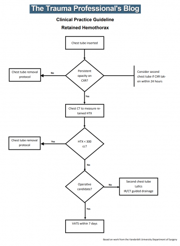
I know it’s small, so just click it to download a pdf copy. I’ve simplified the flow a little as well.

All stable patients with hemothorax admitted to the trauma service were included over a 2.5 year period. The practice guideline was implemented midway through this study period. Before implementation, patients were treated at the discretion of the surgeon. Afterwards, the practice guideline was followed.

Here are the factoids:

  • There were an equal number of patients pre- and post-guideline implementation (326 vs 316)
  • An equal proportion of each group required an initial intervention, generally a chest tube (69% vs 65%)
  • The number of patients requiring an additional intervention (chest tube, VATS, lytics, etc) decreased significantly from 15% to 9%
  • Empyema rate was unchanged at 2.5%
  • Use of VATS decreased significantly from 8% to 3%
  • Use of catheter guided drainage increased significantly from 0.6% to 3%
  • Hospital length of stay was the same, ranging from 4 to 11 days (much shorter than the lytics studies!)

Bottom line: This is how design of practice guidelines is supposed to work. Identify a problem, typically a clinical issue with a large amount of provider care variability. Look at the literature. In general, find it of little help. Design a practical guideline that covers the major issues. Implement, monitor, and analyze. Tweak as necessary based on lessons learned. If you wait for the definitive study to guide you, you’ll be waiting for a long time.

This study did not significantly change outcomes like hospital stay or complications. But it did decrease the number of more invasive procedures and decreased variability of care, with the attendant benefits from both of these. It also dictates more selective (and intelligent) use of additional tubes, catheters, and lytics. 

I like this so much that I’ve incorporated parts of it into the chest tube guideline at my center!

Download the practice guideline here.

Related posts:

Reference: Use of an evidence-based algorithm for patients with traumatic hemothorax reduces need for additional interventions. J Trauma 82(4):728-732, 2017.

Print Friendly, PDF & Email

Surgical Management Of Retained Hemothorax – VATS

I’ve written about the use of lytics to treat retained hemothorax over the past few days. Although it sounds like a good idea, we just don’t know that it works very well. And they certainly don’t work fast. Lengths of stay were on the order of two weeks in both studies reviewed.

The alternative is video assisted thoracoscopic surgery (VATS). So let’s take a look at what we know about it. This procedure is basically laparoscopy of the chest. A camera is inserted, and other ports are added to allow insertion of instruments to suck, peel, and scrape out the hemothorax.

A prospective, multi-center study was performed over a 2 year period starting in 2009. Twenty centers participated, contributing data on 328 patients with retained hemothorax. This was defined as CT confirmation of retained blood and clot after chest tube placement, with evidence of pleural thickening.

Here are the factoids:

  • 41% of patients had antibiotics given for chest tube placement (this is interesting given the lack of consensus regarding their effectiveness!)
  • A third of patients were initially managed with observation, and most of them (82%) did not need any further procedures (83 of 101 patients)
  • Observation was more successful in patients who were older, had smaller hemothoraces (<300cc), smaller chest tubes (!!, <34 Fr), blunt trauma, and peri-procedure antibiotics (?)
  • An additional chest tube was inserted in 19% of patients, image guided drain placement in 5%, and lytics in 5%. Half to two-thirds of these patients required additional management.
  • VATS was used in 34% of patients. One third of them required additional management including another chest tube, another VATS, or even thoracotomy.
  • Thoracotomy was most likely required if there was a diaphragm injury or large hemothorax (<900cc)
  • Empyema and pneumonia were common (27% and 20%, respectively)

Bottom line: There’s a lot of data in this paper. Most notably, many patients resolve their hemothorax without any additional management. But if they don’t, additional tubes, guided drain placement, and lytics work only a third of the time and contribute to additional time in the hospital. Even VATS and thoracotomy require additional maneuvers 20-30% of the time. And infectious complications are common. This is a tough problem!

Tomorrow, I’ll try to roll it all together and suggest an algorithm to try to optimize both outcomes and cost.

Reference: Management of post-traumatic retained hemothorax: A prospective, observational, multicenter AAST study. 72(1):11-24, 2012.

Print Friendly, PDF & Email

Retained Hemothorax Part 2: Lytics (again)

Yesterday, I reviewed a small case report that was published a couple of years ago on lytics for treatment of retained hemothorax. But surely, there must be something better, right?

After digging around, I did find a paper from 2007 that prospectively looked at protocolized management of retained hemothorax, and its aftermath. It was carried out at a busy Level I trauma center over a 16 month period.

All patients with a hemothorax treated with chest tube received daily chest x-rays. Those with significant opacification on day 3 underwent CT scan of the chest. If more than 300 cc of retained blood was present, the patient received streptokinase or urokinase (surgeon preference and drug availability) daily, and rolled around in bed for 4 hours to attempt to distribute it. The chest tube was then unclamped and allowed to drain. This was repeated for 3 days, and if there was still opacification, a repeat CT was obtained. If the volume was still greater than 300 cc, the cycle was repeated for the next 3 days. If the opacification cleared at any point, or the repeat CT showed less than 300 cc, the protocol was stopped and the chest tube removed. If the chest was still opacified after 6 days, VATS was offered.

Here are the factoids:

  • A total of 203 patients with hemothorax were admitted during the study period and 25 (12%) developed a retained hemothorax
  • While a few had treatment start within 4 days, the majority did not receive lytics until day 9 (range 3  –30 days!)
  • The average length of time in hospital after start of lytics was 7 days, leading to a total length of stay of 18 days
  • 92% of patients had “effective” evacuation of their retained hemothorax, although 1 had VATS anyway which found only 100 cc of fluid
  • 16 patients had “complete” evacuation, and 5 had “partial” evacuation
  • There were no hemorrhagic complications, but one third of patients reported significant pain with drug administration

Bottom line: Sounds good, right? The drug seems reasonably effective, although lengths of stay are relatively long. However, streptokinase and urokinase are no longer available in the US, having been replaced with tissue plasminogen activator (tPA). This paper does a cost analysis of lytics vs VATS and found that the former treatment cost about $15000 (drug + hospital stay) vs $34000 for VATS. However, a big part of this was that the drug only cost about $75 per dose. tPA is much more expensive.

So once again, small series, longer lengths of stay, but at least nicely done. Unfortunately, the drug choice is no longer available so use of tPA tilts the balance away from lytics. 

Reference: Intrapleural Thrombolysis for the Management of Undrained Traumatic Hemothorax: A Prospective Observational Study. J Trauma 62(5):1175-1179, 2007.

Print Friendly, PDF & Email

Practice Guideline: Chest Tube Management (Part 2)

Yesterday, I went over the rationale for developing a practice guideline for something as simple and lowly as chest tube management. Today, I’m posting the details of the guideline that’t been in use at my hospital for the past 15 years. I’ve updated it to reflect two lessons learned from actually using it.

Here’s an image of the practice guideline. Click to open a full-size copy in a new window:

Here are some key points:

  • Note the decision tree format. This eliminates uncertainty so that the clinician can stick to the script. There are no hedge words like “consider” used. Just real verbs.
  • We found that hospital length of stay improved when we changed the three parameters from daily monitoring to three consecutive shifts. We are prepared to pull the tube on any shift, not just during the day time. And it also allows this part of the guideline to be nursing driven. They remind the surgeons that criteria are met so we can immediately remove the tube.
  • Water seal is only used if there was an air leak at some point. This allows us to detect a slow ongoing leak that may not be present during our brief inspection of the system on rounds.
  • The American College of Surgeons Committee on Trauma expects trauma centers to monitor compliance with at least some of their guidelines. This one makes it easy for a PI nurse or other personnel to do so.
  • The first of the “new” parts of this guideline is: putting a 7 day cap on failure due to tube output greater than 150cc per three shifts. At that point, the infectious risks of keeping a tube in begin to outweigh its efficacy. Typically, a small effusion may appear the day following removal, then resolves shortly.
  • The second “new” part is moving to VATS early if it is clear that there is visible hemothorax that is not being drained by the system. Some centers may want to try irrigation or lytics, but the data for this is not great. I’ll republish my posts on this over the next two days.

Click here to download a copy of this practice guideline for adults.

Click here to download the pediatric chest tube practice guideline.

Print Friendly, PDF & Email

Practice Guideline: Chest Tube Management (Part 1)

Management of chest tubes is one of those clinical situations that are just perfect for practice guideline development: commonly encountered, with lots of variability between trauma professionals. There are lots of potential areas for variation:

  • How long should the tube stay in?
  • What criteria should be used to determine when to pull it?
  • Water seal or no?
  • When should followup x-rays be done?

Every one of these questions will have a very real impact on that patient’s length of stay and potential for complications.

We developed a chest tube clinical practice guideline (CPG) at Regions Hospital way back in 2004! Of course, there was little literature available to guide us in answering the questions listed above. So we had to use the clinical experience and judgment of the trauma faculty to settle on a protocol that all were comfortable with.

Ultimately, we answered the questions like this:

  • The tube stays in until three specific criteria are met
  • The criteria are: <150 cc drainage over 3 shifts, no air leak, and no residual pneumothorax (or at least a small, stable one)
  • Use of water seal is predicated on whether there was ever an air leak
  • An x-ray is obtained to determine whether any significant pneumo- or hemothorax is present prior to pulling the tube, and 6 hours after pulling it

This CPG has been in effect for over 15 years with excellent results and dramatically shortened lengths of stay.  However, as with any good practice guideline, it needs occasional updates to stay abreast of new research literature or clinical experiences. We recognized that occasional patients had excessive drainage for an extended period of time. This led us to limit the length of time the tube was in to seven days. And we also noted that a few patients had visible hemothorax on their pre-pull imaging. These patients were very likely to return with clinical symptoms of lung entrapment, so we added a decision point to consider VATS at the end of the protocol.

I’ll share the full protocol tomorrow and provide a downloadable copy that you can modify for your own center. I’ll also give a little more commentary on the rationale for the key decision points in this CPG.

Related posts:

Print Friendly, PDF & Email