Category Archives: Thorax

How Safe Is ED Thoracotomy?

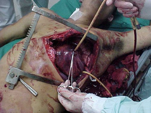
A few weeks ago, I opened a survey to find out common practices regarding performing emergency thoracotomy (EDT) in the emergency department. This procedure is performed at one time or another in most higher level trauma centers. It’s very invasive and is performed in an area that is not really set up for major operative cases. Furthermore, the atmosphere can be chaotic, and stress levels run high.

How safe is this situation? How does personal safety balance out with saving your patient? There are many, many opportunities for injury during this procedure, with significant exposure to blood and other bodily fluids.

A recently published multi-center study examined the potential for exposure during EDT at 16 US trauma centers over a 1.5 year period (14 Level I, 2 Level II). The study was prospective and observational, and was based on questionnaires filled out by all personnel involved in each procedure. A total of 1360 providers submitted information on 305 EDTs.

Here are the factoids:

  • Mechanism was penetrating in 77% of patients, who were predominantly young and male (91%)
  • 15 patients survived (5%), and 4 had residual neurologic impairment
  • Only 56% of respondents wore full personal protective equipment (PPE)
  • There was a 7% exposure rate per EDT(22 incidents), and 1.6% rate per participant in the case
  • The majority of those exposed were trainees (68%) who were injured by something sharp (scalpel 39%, fracture 28%, needle 17%, scissors 3%)
  • There was a strong correlation with PPE use and no exposure during the procedure
  • Only 92% followed their hospital’s occupational exposure protocol if injured (!!!)

Bottom line: Emergency thoracotomy will always be a dangerous procedure. Things happen quickly, there is little time to properly prepare and sharp, pointy things are everywhere. But according to this paper, the actual exposure rate is low. Factoring in the risk of disease transmission, the risk to an individual provider of contracting HIV is 1 in a million, and for hepatitis C is 3 in 100,000

The most distressing part of this study, to me, was the sense of invulnerability of a few of the participants. How can anyone justify not wearing full PPE during an emergency thoracotomy? I believe this represents a very casual attitude toward wearing PPE in any resuscitation. But this study clearly shows a large decrease in exposure rate when full PPEs are worn. Even more disturbing? The fact that 8% chose not to protect themselves by following their own institution’s occupational exposure protocol. Unforgivable!

The main takeaway messages are: always wear your PPE to a trauma resuscitation because you never know when you’ll need to get invasive (and won’t have time to dress up then), and be careful!!

Reference: Occupational exposure during emergency department thoracotomy: A prospective, multi-institution study. J Trauma 85(1):78-84, 2018.

Print Friendly, PDF & Email

Systemic Air Embolism From Chest Trauma

Systemic air embolism (SAE) is an uncommonly encountered but potentially catastrophic complication that trauma professionals will probably see only once or twice in their career. In a couple of very old papers (30-35 years!), it was said to occur in 4 to 14% of patients with severe lung trauma. The pathognomonic finding is complete cardiovascular collapse shortly after intubation and positive pressure ventilation of a patient with significant chest trauma.

SAE can occur after major blunt or penetrating injury. Normally, pressure in the pulmonary arterial and venous branches of the lungs is higher than that of air in the bronchi. If lung parenchyma is torn by gunshot, stab, or blunt pulmonary laceration, then blood can leak into the smaller airways. Unless a major vessel is torn, the volume lost is not of much consequence. A small amount of hemoptysis may be present.

But if positive pressure is applied to the airways, air may be forced into those vascular structures. If the injury involves a pulmonary vein, the gas bubbles enter the left heart and and then the systemic circulation. Only a cc or two of air in the cerebral circulation or coronary arteries can cause a rapidly fatal condition.

How can a trauma professional suspect that a patient may be susceptible to SAE? Look for evidence of hemoptysis in patients with penetrating chest injury or severe blunt chest trauma. Unfortunately, this occurs in only a few patients and its absence is not helpful.

Always suspect SAE if your patient suffers circulatory collapse or arrest shortly after intubation and positive pressure ventilation. This is especially true in patients who were very stable up until that point. Ultrasound may be used to detect air bubbles in the left heart. Transesophageal echo is even better, but not readily available in the ED.

What can be done if your patient (nearly) arrests after intubation and you suspect SAE? The recommended treatment is single lung ventilation and thoracotomy on the injured side. The injured side is obvious in patients with unilateral penetrating injury, but much more difficult to determine in blunt trauma where either lung may be involved. A quick chest x-ray could be obtained, but may not localize the injured lung.

If the left lung is involved, push the endotracheal tube into the right mainstem bronchus to eliminate the abnormal pressure gradient in the injured lung. If the right lung in injured, a dual lumen endotracheal tube should be inserted for single-lung ventilation. Unfortunately, this requires fiberoptic tools and is not available in the field or most emergency departments.

If single-lung ventilation can be accomplished, this may buy some time to try to resuscitate your patient. A thoracotomy of the injured side can also be carried out to occlude the hilum of the lung in an attempt to stop any further embolism. Initial clamping should be carried out by hand, as using a crushing clamp commits the patient to an emergency pneumonectomy (if they survive beyond this point).

Overall survival is dismal. Old data suggests that more than 80% of blunt trauma victims with SAE die, as well as half of patients with penetrating injury. These numbers are even lower once the patient arrests. The key to survival is avoiding unnecessary intubation in patients with potential SAE, and moving to single-lung ventilation quickly in those who have developed it.

Print Friendly, PDF & Email

Flying Or Diving After Traumatic Pneumothorax: Part 2

Yesterday, I wrote about the accepted management of and delay in flying due to traumatic pneumothorax. I republished the post because of the very recent acceptance for publication of a paper from Oregon Health Science University in Portland. The authors specifically tried to assess timing of chest tube removal and long-distance flight, and to measure the risk of pneumothorax recurrence or other complications.

The authors performed a retrospective review of a series of military patients who had sustained chest injuries that were treated with chest tubes over a 5 year period from 2008 to 2012. After tube removal and a pneumothorax-free period of at least 24 hours (by chest x-ray), the patients were then transported by air from the military theater back to the United States.

Here are the factoids:

  • Of 517 patients screened in the military trauma registry database, only 73 were available for study after applying exclusion criteria
  • Subjects were predominantly young and male, as one would expect from the injured military population, and 74% were injured by a penetrating mechanism
  • Median time that the chest tube was in place was 4 days, and median time from tube removal to flight was 2.5 days
  • All patients had post-flight documentation available for review, but only half (37) had in-flight documentation available
  • Nearly half (40%) had positive pressure ventilation in place during the flight
  • Five patients had “in-flight medical concerns” (4 were ventilated), but none were related to the pneumothorax. The four ventilated patients had ventilator issues, the non-vented patient had “self-limited discomfort without evidence of respiratory distress.”
  • None of the subjects developed a recurrent pneumothorax, either post-flight or over the following 30 days

The authors conclude that air travel after tube removal and a 24-72 hour observation period “appears safe.”

Bottom line: Not so fast! This is yet another small, retrospective study making grand claims. The study group is a very unique population: healthy, fit young men with penetrating injury. Your average civilian trauma patient is older, less healthy, and usually has a blunt mechanism with multiple rib fractures. In-flight documentation was not available in half of the cases. And a full medical team was present on the aircraft had a problem actually occurred.

Contrast this with a civilian patient on a commercial aircraft with very limited medical equipment and expertise on board. What could go wrong? I definitely do not recommend changing our practice on these patients yet based on this one paper. Until we have better guidance (more good papers) stick to the usual wait time to ensure a safe flight for your patient.

Reference: Trauma patients are safe to fly 72 hours after tube thoracostomy removal. J Trauma, published ahead of print, May 18 2018.

Print Friendly, PDF & Email

Flying Or Diving After Traumatic Pneumothorax: Part 1

Today, I’m dusting off an old post on flying and diving after pneumothorax. This shows the thinking up until last year. Tomorrow, I’ll write about a new paper that suggests that we can shorten the “no-fly” time considerably.

Hint: no changes to the diving recommendations. One pneumothorax is likely to ground you forever.

Patients who have sustained a traumatic pneumothorax occasionally ask how soon they can fly in an airplane or scuba dive after they are discharged. What’s the right answer?

The basic problem has to do with Boyle’s Law (remember that from high school?). The volume of a gas varies inversely with the barometric pressure. So the lower the pressure, the larger a volume of gas becomes. Most of us hang out pretty close to sea level, so this is not an issue. But for flyers or divers, it may be.

Flying

Helicopters typically fly only one to two thousand feet above the ground, so the air pressure is about the same as standing on the earth. However, flying in a commercial airliner is different. Even though the aircraft may cruise at 30,000+ feet, the inside of the cabin remains considerably lower though not at sea level. Typically, the cabin altitude goes up to about 8,000 to 9,000 feet. Using Boyle’s law, any volume of gas (say, a pneumothorax in your chest), will increase by about a third on a commercial flight.

The physiologic effect of this increase depends upon the patient. If they are young and fit, they may never know anything is happening. But if they are elderly and/or have a limited pulmonary reserve, it may compromise enough lung function to make them symptomatic. And having a medical problem in an aluminum tube at 30,000 feet is never good.

Commercial guidelines for travel after pneumothorax range from 2-6 weeks. The Aerospace Medical Association published guidelines that state that 2-3 weeks is acceptable. The Orlando Regional Medical Center reviewed the literature and devised a practice guideline that has a single Level 2 recommendation that commercial air travel is safe 2 weeks after resolution of the pneumothorax, and that a chest x-ray should be obtained immediately prior to travel to confirm resolution.

Diving

Diving would seem to be pretty safe, right? Any pneumothorax would just shrink while the diver was at depth, then re-expand to the original size when he or she surfaces, right?

Not so fast. You are forgetting why the pneumothorax was there in the first place. The lung was injured, most likely via tearing it, penetration by something sharp, or popping a bleb. If the injured area has not completely healed, then air may begin to escape through it again. And since the air used in scuba diving is delivered under pressure, this could result in a tension pneumothorax.  This is disastrous underwater!

Most injuries leading to pneumothorax heal completely. However, if there are bone spicules stuck in the lung or more complicated parenchymal injuries from penetrating injury, they may never completely heal. This makes the diver susceptible to a tension pneumothorax anytime they use their regulator.

Bottom line: Most patients can safely travel on commercial aircraft 2 weeks after resolution of pneumothorax. Ideally, a chest xray should be obtained shortly before travel to confirm that it is gone. Helicopter travel is okay at any time, since they typically fly at 1,500 feet or less.

Divers should see a physician trained in dive medicine to evaluate their injury and imaging prior to making another dive.

Tomorrow: new info on flying after pneumothorax

References:

  • Divers Alert Network – Pneumothorax – click to download
  • Practice Guideline, Orlando Regional Medical Center. Air travel following traumatic pneumothorax. October 2009.
  • Medical Guidelines for Airline Travel, 2nd edition. Aerospace Medical Association. Aviation, Space, and Environmental Medicine 74(5) Section II Supplement, May 2003.
Print Friendly, PDF & Email

Off-Label Use of the Foley (Urinary) Catheter

Foley catheters are a mainstay of medical care in patients who need control or measurement of urine output. Leave it to trauma surgeons to find warped, new ways to use them!

Use of these catheters to tamponade penetrating cardiac injuries has been recognized for decades (see picture, 2 holes = 2 catheters!). Less well appreciated is their use to stop bleeding from other penetrating wounds.

Foley catheters can be inserted into just about any small penetrating wound with bleeding that does not respond to direct pressure. (Remember, direct pressure is applied by one or two fingers only, with no flat dressings underneath to diffuse the pressure). Arterial bleeding, venous bleeding or both can be controlled with this technique.

In general, the largest catheter with the largest possible balloon should be selected. It is then inserted directly into the wound until the entire balloon is inside the body. Inflate the balloon using saline until firm resistance is encounted, and the bleeding hopefully stops. Important: be sure to clamp the end of the catheter so the bleeding doesn’t find the easy way out!

Use of catheter tamponade buys some time, but these patients need to be in the OR. In general, once other life threatening issues are dealt with in the resuscitation room, the patient should be moved directly to the operating room. In rare cases, an angiogram may be needed to help determine the type of repair. However, in the vast majority of cases, the surgeon will know exactly where the injury is and further study is not needed. The catheter is then prepped along with most of the patient so that the operative repair can be completed.

Tomorrow: an off-label use for this catheter in abdominal trauma!

Print Friendly, PDF & Email