Category Archives: Thorax

Chest Tubes: Size Doesn’t Matter – Part 2

A few days ago, I wrote about a paper that seemed to suggest that using a smaller chest tube (28-32 Fr) vs larger ones (36-40 Fr). The results suggested that their function was very similar. I emphasized that I thought the result was intriguing, because I’m of the opinion that bigger is better for getting clotted blood out. However, I am amenable to changing my mind based on newer, better data.

But I did caution readers that I would like to see more data. One study should never change your practice! Then I see a lot of chatter on Twitter about another study from 2016 that looks at even smaller tubes, with people saying they will now switch to pigtail catheters (12 Fr)!!

First, not a logical progression of thinking there. And second, let’s take an actual look at the paper. It’s from an emergency medicine group in Fukui, Japan, which retrospectively reviewed their 7 year experience with using a small (20-22 Fr) vs large (28 Fr) tubes. They identified a total of 124 chest tube insertions to compare, 68 small and 56 large.

Now let’s look at the factoids:

  • Demographics, mechanism, and ISS were the same between groups
  • Duration of insertion and initial drainage were also the same between groups
  • Complication rates were similar, with 1 empyema and 2 retained hemothoraces in each group
  • Additional tubes were place in 2 patients with small tubes vs 4 with large tubes
  • Thoracotomy was performed in 2 patients with small tubes vs 1 with a large tube

Based on all of this, the authors concluded that there was no difference in drainage efficacy, complications, or need for additional invasive procedures.

Wait a minute!! Again, if you only read the abstract, you might be led to start using ever smaller chest tubes. But read the entire paper! There are many problems with this paper, including:

  • It’s a very small, retrospective review. This automatically means that the statistical power is suspect.
  • Why did they only document 124 insertions over 7 years?? That’s about one every 3 weeks! Either a lot of data are missing or they are not very busy. But Fukui Prefectural Hospital has over 1000 beds! So it’s the former, not the latter.
  • The retrospective nature means it is not possible to determine why a particular tube size was chosen. Roll of the dice? This fact alone introduces a huge potential for selection bias. Was a smaller tube selected because the hemothorax looked smaller? Probably! The fact that 4 patients with larger tubes had another one placed suggests that they were being used for larger collections. And patients with higher ISS tended to get bigger tubes.

Bottom line: Don’t change your practice based on this paper. And certainly don’t choose to use even smaller pigtails. And of course, always critically read any paper that you like to make sure you are not cherry picking the ones you choose to believe. IMHO, it’s still best to use big (36 Fr) or bigger (40 Fr).

Reference: Small tube thoracostomy (20-22 Fr) in emergent management of chest trauma. Injury 48:1884-1887, 2016.

Print Friendly, PDF & Email

Chest Tube Size Doesn’t Matter?

It’s great when you read a study that supports your own biases. But it’s not pleasant at all when you find one that refutes what you’ve been teaching for years. Well, I found one of those and I wanted to share it with you.

I’ve always said that there are only two sizes of chest tube for trauma, big (36Fr) and bigger (40Fr). Although there was never any good literature, it seemed intuitive that a large tube would help ensure drainage of bigger clots if hemothorax was present.

A multicenter observational study was carried out that looked at 353 chest tube insertions. This work monitored retained hemothorax or pneumothorax, the need for tube reinsertion or invasive procedure due to incomplete drainage, and pain during insertion.

Here are the factoids:

  • There was roughly a 50:50 large (36-40Fr) vs small (28-32Fr) mix of chest tubes
  • Tubes inserted for hemothorax were also a 50:50 mix of large vs small
  • The initial amount of blood out was small and about the same for both groups
  • There was no significant difference in pneumonia, retained hemothorax, or empyema
  • The need for an invasive procedure (VATS or thoracotomy) was about 11% in both groups
  • Interestingly, there was no difference in visual analog pain score between the groups either.

Bottom line: Basically, large tube and small tube were the same. So maybe chest tube size selection doesn’t matter as much as we (I?) think. It seems to make sense to select a tube size based on your patient’s chest wall, not dogma. Although subjective pain seems to be the same as well, pain and sedation management are key because this is not a fun procedure for the patient, regardless of tube size. I’m not fully convinced yet, and would like to see an additional confirmation study if possible.

Reference: Does size matter? A prospective analysis of 28–32 versus 36–40 French chest tube size in trauma. J Trauma 72(2):422-427, 2012.

Print Friendly, PDF & Email

How To: Insert A Small Percutaneous Chest Tube

This short (10 minute) video demonstrated the technique for inserting small chest tubes, also known as “pigtail catheters.” It features Jessie Nelson MD from the Regions Hospital Department of Emergency Medicine. It was first shown at the third annual Trauma Education: The Next Education conference in September 2015, for which she was a course director.

Please feel free to leave any comments or ask any questions that you may have.

YouTube player

Related posts:
Pigtail catheters vs regular chest tubes
Tips for regular chest tubes 

Print Friendly, PDF & Email

How To Evaluate A Stab To The Diaphragm – Part 2

Yesterday I gave a little perspective on the use of CT in assessing the diaphragm after penetrating injury. Today, I’ll break it down into some practical steps you can follow the next time you see one.

Step 1. Stable or unstable? If your patient arrives with unstable vital signs, and there is no other source but the abdomen, the answer is simple. Go to the OR for a laparotomy. Period. They are exsanguinating and the hemorrhage needs to be stopped.

Step 2. Mark the sites of penetration and take a chest x-ray. This will let you evaluate the potential trajectory of the object, and will give you your first glimpse of the diaphragm.

Step 3. Examine the abdomen. Actually, you should be doing this at the same time you are setting up for Step 2. If your patient has peritoneal signs, no further evaluation is needed. Just go to the OR for laparotomy. Look at the chest x-ray once you get there.

Step 4. Right side? If your appreciation of the path of penetration involves just the liver, take the patient to CT for evaluation of chest, abdomen, and pelvis. You need to see all three of these areas to assess for blood and fluid in both body cavities. After the study, if you still think the injury is limited to the liver, admit the patient for observation.

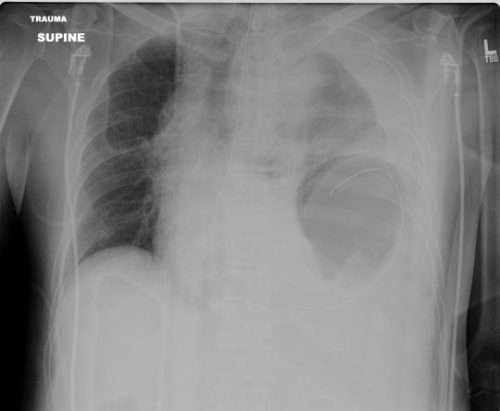
Step 5. Left side? Look at that chest x-ray again. If there are any irregularities at all, strongly consider going to the OR and starting with diagnostic laparoscopy. These irregularities can be glaring, like in the x-ray above. But they can be subtle, like some haziness above the diaphragm or small hemothorax. Obviously, if the injury is as clear as on the x-ray above, just open the abdomen. But if in doubt, start small. And remember my advice on “lunchothorax.”

Step 6. Admit and observe. Check the abdomen periodically, and repeat the chest x-ray daily. If anything changes, consider diagnostic laparoscopy. As a general rule, I don’t keep patients NPO “just in case.” Most will pass this test, and I don’t see a reason to starve my patients for the low likelihood they need to go to the OR.

Step 7. Make sure your patient gets a follow up evaluation. See them in your outpatient clinic, get a final chest x-ray and abdominal exam before you completely clear them.

Print Friendly, PDF & Email