Category Archives: Head

Best Of AAST #4: TBI and Antiplatelet / Antithrombotic Agents

More and more people are taking antiplatelet or antithrombotic agents for a variety of medical conditions. One of the dreaded side effects of these medications is undesirable bleeding, particularly after injury. This is especially true if the bleeding occurs inside the skull after any kind of head trauma.

Which agents, if any, lead to worse outcomes? The literature has been a bit inconsistent over the past 10 years. A group from HCA Healthcare reviewed the trauma registries from 90 hospitals, which I presume are in the HCA system. They included patients patients who suffered a ground level fall and were 65 years or older. They excluded those who had a significant injury to regions other than the head.

Here are the factoids:

  • Over, 33,000 patient records were reviewed, with an average age of 81
  • Nearly half were on single or multiple anti-thrombotic therapy (!)
  • The proportion of patients sustaining a “TBI” was roughly the same (21%) whether they were not on anti-thrombotic therapy or not
  • Apixaban and rivaroxiban were associated with lower rates of “TBI” (13-16%)
  • Clopidogrel was associated with a higher “TBI” rate (23%)
  • Patients requiring brain surgery  were more common in patients taking aspirin plus clopidogrel (2.9%) vs all the others (2%) and this was statistically significant
  • None of the treatment regimens were associated with higher mortality (roughly 2-3%)
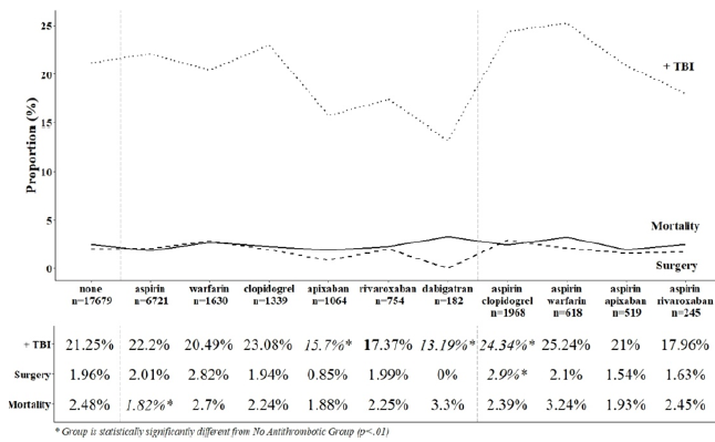
The authors conclude that anti-thrombotic use in the elderly who suffer a ground level fall are not at risk for increased mortality and that they may have negligible impact on management.

My comments: The one thing that makes this abstract difficult to read is their use of the term TBI, which is why I put it in quotes above. I think that the authors are conflating this acronym with intracranial hemorrhage. It’s a bit confusing, because I think of TBI as a term that means the head was struck and either left a physical mark (bump on the outside or blood on the inside) or there was known or suspected loss of consciousness. They are apparently using  it to describe intracranial bleeding seen on CT.

And because this is a registry study, many of the patient-specific outcome details cannot be analyzed. Mortality and operative rates are very crude outcomes. What about some of the softer ones? Although the average GCS was stated to be 14.5, it would be interesting to know how many of these patients were able to return to their previous living situation, and how many were significantly impaired even though they didn’t die or need an operation.

Here are my questions for the presenter and authors:

  • How do you define a TBI in this study? Could it be just a concussion? Does it require some type of blood in the head? Assuming that there are lots of TBIs that occur without intracranial bleeding, including such patients in your analyses will skew the data toward lower incidence and will dilute out the patients with hemorrhage.
  • What was the length of your study? If it includes data that is older than six years or so, it may under-represent the use of some of the direct oral anticoagulant drugs (DOACs).
  • Are half of your elderly falls patients really on anti-thrombotic therapy? This is a shocking number, and seems to be high in my experience. Since your study was distributed across a large number of hospitals, it brings up the question of whether so many of our elders really need this medication.
  • Do you have any sense for how your various subgroups fared in terms of their discharge disposition? You conclude that the use of anti-thrombotic agents isn’t so bad, really. At least when it comes to needing brain surgery or dying. But are there other cognitive issues that are common that might encourage trauma professionals to continue to look at these drugs with a wary eye?

This is important work, and I am anticipating a great discussion after your presentation.

Reference: Antiplatelet and antiplatelet agents, alone and in combination, have minimal impact on traumatic brain injury (TBI) incidence, need for surgery, and mortality in ground level falls (GLFs): a multi-institutional analysis of 33,710 patients. AAST 2020 Oral Abstract # 7.
More On CSF Rhinorrhea/Otorrhea


Trauma professionals worry about stuff. Like just about everything, really. Sometimes we have good guidance (research) to help us decide what to do. Many times, we don’t. Management of rhinorrhea and otorrhea from CSF leak after trauma is definitely one of those things.

I’ve seen a variety of treatments used in these patients over the years. Is it really a CSF leak? Let’s get a beta-2 transferrin test (see below). Can’t the patient get meningitis? Their may be concomitant sinus fracture and bacterial contamination, so why not give antibiotics? Or vaccinate them?

The Cochrane library contains a vast number of reviews of common clinical questions. One of those questions just happens to be the utility of giving prophylactic antibiotics in patients with basilar skull fracture. Interestingly, they’ve been reviewing and re-reviewing this question about every 5 years, since 2006. During the three reviews done, there have been no additional research papers published on the topic.

Here are the factoids:

  • Studies that specifically examined the use of prophylactic antibiotics in patients with basilar skull fracture were reviewed. All included meningitis as one of the outcome parameters.
  • There were only 5 high quality (randomized, controlled) trials, with a total of 208 participants
  • There were an additional 17 lower quality trials published, but no conclusions could be reached from them due to methodology problems
  • In the high quality trials, there were no differences in the incidence of meningitis, mortality, or meningitis-related mortality
  • There were no specific adverse effects related to antibiotic administration. But one of the high quality studies did note a shift to higher counts of pathologic bacteria in the posterior nasopharynx in the antibiotic group.
  • No studies on the use of meningitis vaccinations exist. A survey of UK physicians showed that 35% recommend at least one vaccine, typically for Strep Pneumo.

Bottom line: There is still no good evidence to support the use of prophylactic antibiotics or meningitis vaccination in patients with CSF leak from uncomplicated basilar skull fracture. When you see surveys that show some physicians promoting a treatment and others doing nothing, it means there is most likely no significant benefit. If there were a big difference, we would have seen it by now! And giving drugs (antibiotics, vaccines) that have no proven use is expensive and can always lead to unexpected complications. 

References:

  • Immunisations and antibiotics in patients with anterior skull base cerebrospinal fluid leaks. J Laryngol Otol 128(7):626-629, 2014.
  • Antibiotic prophylaxis for preventing meningitis in patients with basilar skull fractures. Cochrane Database Syst Rev April 28, 2015.

Tips For Taking Care Of CSF Leaks

The management of CSF leaks after trauma remains somewhat controversial. The literature is sparse, and generally consists of observational studies. However, some general guidelines are supported by large numbers of retrospectively reviewed patients.

  • Ensure that the patient actually has a CSF leak. In most patients, this is obvious because they have clear fluid leaking from ear or nose that was not present preinjury. Here are the options when the diagnosis is less obvious (i.e. serosanguinous drainage):
    • The “halo” or “double ring sign” is a form of pillow chromatography. The blood components separate from the CSF as they move through the pillow fabric, creating a clear ring or halo surrounding a bloody spot. This is the cheapest, fastest test and is actually fairly reliable.
    • High resolution images of the temporal bones and skull base. If an obvious breach is noted, especially if fluid is seen in the adjacent sinuses, then a CSF leak is extremely likely.
    • Glucose testing. CSF glucose is low compared to serum glucose.
    • Beta 2 transferrin assay. Don’t do it!! This marker is very specific to CSF. However, the test is expensive and results may take several days to a few weeks to receive. Most leaks will have closed before the results are available, making this a poor test.
  • Place the patient at bed rest with the head elevated. The basic concept is to decrease intracranial pressure, which in turn should decrease the rate of leakage. This same technique is used for management of mild ICP increases after head injury.
  • Consider prophylactic antibiotics carefully. The clinician must balance the likelihood of meningitis with the possibility of selecting resistant bacteria. If the likelihood of contamination is low and the patient is immunocompetent, antibiotics may not be needed.
  • Ear drops are probably not necessary. They may confuse the picture when gauging resolution of the CSF leak.
  • Wait. Most tramatic leaks will close spontaneously within 7-10 days. If it does not, a neurosurgeon or ENT surgeon should be consulted to consider surgical closure.

References:

  1. Brodie HA, Thompson TC. Management of complications from 820 temporal bone fractures. Am J Otol, 1997;18:188-197.
  2. Brodie HA. Prophylactic antibiotics for posttraumatic cerebrospinal fluid fistulas. Arch Otolaryngol Head, Neck Surg. 123:749-752.

Management Of Blunt Carotid / Vertebral Injury

Yesterday I reviewed the most commonly used grading system for blunt carotid / vertebral injury (BCVI). Today, I’ll describe the usual management of these injuries, by grade. Unfortunately, there is a paucity of definitive literature to guide us because these injuries are rare. So here are our best guesses to date.

There are basically three modalities at our disposal for managing BCVI: antithrombotic medication (heparin and/or antiplatelet agents), surgery, and therapeutic angiographic procedures. The choice of therapy is usually based on surgical accessibility and patient safety for anticoagulation. We do know that a number of studies have shown a decrease in stroke events in patients who are heparinized. Unfortunately, this is not always possible due to associated injuries. Antiplatelet agents are usually tolerated after acute trauma, especially low-dose aspirin. Several studies have shown little difference in outcomes in patients receiving heparin vs aspirin/clopidogrel for BCVI.

So what to do? Here are some broad guidelines:

  • Grade I (intimal flap). Heparin or antiplatelet agents should be given. If heparin can be safely administered, it may be preferable in patients who will need other surgical procedures since it can be rapidly reversed just by stopping the infusion. These lesions generally heal completely, so a followup CT angiogram should be scheduled in 1-2 weeks. Medication can be stopped when the lesion heals.
  • Grade II (flap/dissection/hematoma). These injuries are more likely to progress, so heparin is preferred if it can be safely given. Stenting should be considered, especially if the lesion progresses. Long-term anti-platelet medication may be required.
  • Grade III (pseudoaneurysm). Initial heparin therapy is preferred unless contraindicated. Stable pseudoaneurysms should be followed with CTA every 6 months. If the lesion enlarges, then surgical repair should be carried out in accessible injuries, or stenting in inaccessible ones.
  • Grade IV (occlusion). Heparin therapy should be initiated unless contraindicated. Patients who do not suffer a catastrophic stroke may do well with followup antithrombotic therapy. Endovascular treatment does not appear to be helpful.
  • Grade V (transection with extravasation). This lesion is frequently fatal, and the bleeding must be addressed using the best available technique. For lesions that are surgically accessible, the patient should undergo the appropriate vascular procedure. Inaccessible injuries should undergo angiographic treatment, and may require embolization to control bleeding without regard for the possibility of stroke.

References:

  • Scott WW, Sharp S, Figueroa SA, et al. Clinical and radiographic outcomes following traumatic Grade 1 and 2 carotid artery injuries: a 10-year retrospective analysis from a Level I trauma center. J Neurosurg 122:1196, 2015.
  • Scott WW, Sharp S, Figueroa SA, et al. Clinical and radiographic outcomes following traumatic Grade 3 and 4 carotid artery injuries: a 10-year retrospective analysis from a Level 1 trauma center. J Neurosurg 122:610, 2015.
  • Scott WW, Sharp S, Figueroa SA, et al. Clinical and radiological outcomes following traumatic Grade 1 and 2 vertebral artery injuries: a 10-year retrospective analysis from a Level 1 trauma center. J Neurosurg 121:450, 2015.
  • Scott WW, Sharp S, Figueroa SA, et al. Clinical and radiological outcomes following traumatic Grade 3 and 4 vertebral artery injuries: a 10-year retrospective analysis from a Level I trauma center. The Parkland Carotid and Vertebral Artery Injury Survey. J Neurosurg 122:1202, 2015.


What Does Blunt Carotid / Vertebral Injury Look Like?

In my last two posts, I reviewed who is at risk for blunt carotid / vertebral injury (BCVI) and how to find it. But what does it actually look like, and how is it classified?

A seminal paper from Denver Health (aka Denver General Hospital) in 1999 proposed the most commonly used grading system for BCVI. This Denver scale should not be confused with the Denver criteria that predict risk for BCVI. Here’s a nice graphic that explains the classifications:

Grade I: A mild intimal irregularity is seen. Note the abnormal  narrowed area, representing a small intimal injury, possibly with a small amount of clot.

Grade II: This grade has several presentations. There may be a intraluminal thrombosis/hematoma with (left) or without (right) an intimal flap, or a flap alone (center)

Grade III: There is a full-thickness injury to the vessel with a contained extraluminal extravasation (pseudoaneurysm)

Grade IV: The vessel is completely occluded by flap or thrombus

Grade V: The artery is transected and freely extravasating

In the next post, I’ll finish off with a summary of the treatments for these injuries.

Reference: Blunt carotid arterial injuries: implications of a new grading scale. J Trauma. 47(5): 845-53, 1999.