Category Archives: Head

Everything You Wanted To Know About: Cranial Bone Flaps

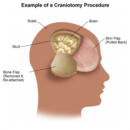
Patients with severe TBI frequently undergo surgical procedures to remove clot or decompress the brain. Most of the time, they undergo a craniotomy, in which a bone flap is raised temporarily and then replaced at the end of the procedure.

But in decompressive surgery, the bone flap cannot be replaced because doing so may increase intracranial pressure. What to do with it?

There are four options:

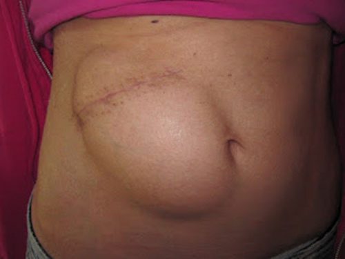
  1. The piece of bone can buried in the subcutaneous tissue of the abdominal wall. The advantage is that it can’t get lost. Cosmetically, it looks odd, but so does having a bone flap missing from the side of your head. And this technique can’t be used as easily if the patient has had prior abdominal surgery.

2. Some centers have buried the flap in the subgaleal tissues of the scalp on the opposite side of the skull. The few papers on this technique demonstrated a low infection rate. The advantage is that only one surgical field is necessary at the time the flap is replaced. However, the cosmetic disadvantage before the flap is replaced is much more pronounced.

3. Most commonly, the flap is frozen and “banked” for later replacement. There are reports of some mineral loss from the flap after replacement, and occasional infection. And occasionally the entire piece is misplaced. Another disadvantage is that if the patient moves away or presents to another hospital for flap replacement, the logistics of transferring a frozen piece of bone are very challenging.

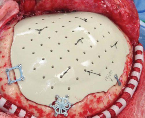
4. Some centers just throw the bone flap away. This necessitates replacing it with some other material like metal or plastic. This tends to be more complicated and expensive, since the replacement needs to be sculpted to fit the existing gap.

So which flap management technique is best? Unfortunately, we don’t know yet, and probably never will. Your neurosurgeons will have their favorite technique, and that will ultimately be the option of choice.

Reference: Bone flap management in neurosurgery. Rev Neuroscience 17(2):133-137, 2009.

Print Friendly, PDF & Email

Sports Drinks And Electrolyte Replacement In TBI

Yesterday, I wrote about the (lack of) effectiveness of forcing hypernatremia in the management of TBI. However, we do know that some of our head injured patients have trouble maintaining a normal sodium level, and if it drops quickly or too far, hyponatremia can certainly cause problems. Trauma professionals have a number of tools to help fix this, including salt supplements or tablets, saline infusions, or even hypertonic saline in more difficult cases.

But what about using a sports drink to replace electrolytes? Isn’t that what athletes do? There are quite a few of these sports drinks on the market, and new ones seem to appear every week. Common examples are Gatorade, Powerade, Muscle Milk, Vitamin Water, 10-K Thirst Quencher, and many more. What if your brain injured patients eschews the salt tabs and insists on pounding down sports drinks all day?

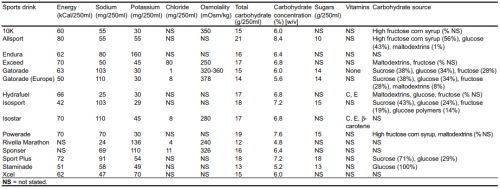
Here is a table from an old sports medicine paper that describes the composition of a number of sports drinks from back in the day. Some, like Gatorade, are still around. (Click image to see a bigger, readable version)

Note that the electrolyte results are in mg/250cc, so I will translate to meq/liter for you. Gatorade had the highest sodium concentration at the time, 20meq/L, and one of the lowest potassiums at 3meq/L. The majority of the current day sports drinks have about the same electrolyte composition. Note that they are all a bit hyperosmolar (300+ mOsm), and this is made possible by added carbohydrate from some type of sugar. The carb is usually in the form of sucrose, dextrose, and/or high fructose corn syrup (yum!).

Bottom line: Your typical sports drink is equivalent to D30 in 0.1 normal saline. Not good for your TBI patient when consumed for sodium supplementation. It will actually drive the serum sodium down when consumed in quantity. Make sure your patients steer clear of this stuff until their brain injury is healed and they are running their next marathon.

Reference: The Effectiveness of Commercially Available Sports Drinks. Sports Med 29(3):181-209, 2000.

Print Friendly, PDF & Email

Targeted Hypernatremia In Trauma Brain Injury: Does This Work?

Traumatic brain injury (TBI) frightens and confuses most trauma professionals. The brain and its workings are a mystery, and there is very little real science behind a lot of what we do for TBI. One thing that we do know is that intracranial hypertension is bad. And another is that we do have some potent drugs (mannitol, hypertonic saline) to treat it emergently.

So if we can “dry out” the brain tissue on a moment’s notice and drop the ICP a bit with a hit of sodium, doesn’t it stand to reason that elevating the sodium level constantly might keep the brain from becoming edematous in the first place? Many neurosurgeons buy into this, and have developed protocols to maintain serum sodium levels in the mid-140s and higher. But what about the science?

A nice review was published in Neurocritical care which identified the 3 (!) papers that have promoted this practice in humans with TBI. In general, there was a decrease in ICP in the patients in the cited papers. Unfortunately, there were also a number of serious and sometimes fatal complications, including pulmonary edema and renal failure requiring hemodialysis. These complications generally correlated with the degree of hypernatremia induced. Papers were also reviewed that involved patients with other brain injury, not caused by trauma. Results were similar.

Bottom line: There is no good literature support, standard of care, or even consensus opinion for prophylactically inducing hypernatremia in patients with TBI. The little literature there is involves patients with severe TBI and ICP monitors in place. There is nothing written yet that justifies the expense (ICU level care) and patient discomfort (frequent blood draws) of using this therapy in patients with milder brain injury and a reliable physical exam. If you want to try out this relatively untried therapy, do us all a favor and design a nice study to show that the benefits truly outweigh the risks. 

And if you can point me to some supportive literature that I’ve missed, please do so!

Related posts:

References:

  • Induced and sustained hypernatremia for the prevention and treatment of cerebral edema following brain injury. Neurocrit Care 19:222-231, 2013.
  • Continuous hyperosmolar therapy for traumatic brain injury-induced cerebral edema: as good as it gets, or an iatrogenic secondary insult? J Clin Neurosci 20:30-31, 2013.
  • Continuous hypertonic saline therapy and the occurrence of complications in neurocritically ill patients. Crit Care Med 37(4):1433-1441, 2009. -> Letter to the editor Crit Care Med 37(8):2490-2491, 2009.
Print Friendly, PDF & Email

A Blood Test For TBI? Part 3

The FDA announced approval of a blood test that incorporates both GFAP and UCH-L1. Approval was based on two as yet to be published studeis titled Evaluation of Biomarkers of Traumatic Brain Injury (ALERT-TBI) and Evaluation of Biomarkers of Traumatic Brain Injury Extension Study (ALERT-TBIx), and passed after less than 6 months of evaluation. Yes, more silly acronyms, I know.

The studies were designed to “evaluate the utility of the Banyan UCH-L1/GFAP Detection Assay as an aid in the evaluation of suspected traumatic brain injury (Glasgow Coma Scale score 9-15) in conjunction with other clinical information within 12 hours of injury to assist in determining the need for a CT scan of the head.”

The former study started in 2012 and involved 2011 patients! The latter had only 119 patients, starting in 2015. Now, I have no access to their data, so I can’t tell what the FDA saw.

From Banyan Biomarkers’ website:

“The CT scan is widely available to assist clinicians in the evaluation of TBI, however, CT scans do not provide a clear and objective answer and scanning may increase the risk for radiation-induced cancer. Furthermore, over 90% of patients presenting to the emergency department with mild TBI, sometimes described as “concussion”, have a negative CT scan. Despite these limitations, nearly all patients are sent for a CT, which results in increased costs to the healthcare system and unnecessary patient exposure to radiation.”

Here are the (very) few factoids that I can find:

  • CT scan results were compared to the Brain Trauma Indicator (BTI) blood test (GFAP + UCH-L1)
  • BTI predicted a positive CT scan 98% of the time
  • It predicted a negative CT scan 99.6% of the time
  • Time to process the test is currently 4 hours

Bottom line: Sounds promising, right? Based on the data summarized over the last two days, I wouldn’t be too excited about this test, but the FDA was able to look at a study that I can’t. It appears that the negative predictive value is excellent, so I can see the application.

That being said, 4 hours is way to long. We can’t have a patient sitting in the ED waiting for the results to come back to decide whether they need a head CT. And how long will it take the assay to be widely available?

The devil will be in the details. What types of intracranial lesions were detected. Are the negative predictive values the same for subarachnoid, subdural, epidural, or intraparenchymal bleeds? And finally, how expensive will it be? How does the cost for the test compare to the cost of a CT scan done in 5 minutes?

I’ll let you know more as the details emerge. But don’t look for, or plan to use, this test at your hospital any time soon. There’s more work to do!

Reference: Banyan Biomarkers (banyanbio.com)

Print Friendly, PDF & Email

A Blood Test For TBI? Part 2

Yesterday, I wrote about one blood biomarker, GFAP, and its possible application in detecting traumatic brain injury (TBI). Today, I’ll discuss a complementary marker called UCH-L1.

Fewer studies have been done looking at the utility of UCH-L1 in detecting TBI than of GFAP.  A review article published last month pooled existing literature to get a sense of how good this biomarker really is. It also examined the risk of bias due to the small numbers of studies involved.

Here are the factoids:

  • Only 38 abstracts were eligible, but full text was available for analysis in only 13 (meaning it was only an abstract and never passed muster for publication). The authors of the published studies were contacted for additional information, which is an interesting (and helpful) practice.
  • Of all of those, only 4 were selected for meta-analysis! This significantly limited the value of the analysis.
  • Serum UCH-L1 has a high accuracy in predicting CT findings in mild to moderate TBI, but there is a high risk of bias affecting this result
  • Plasma UCH-L1 has a moderate accuracy predicting CT findings across all GCS levels, with a low risk of bias
  • Pooling all studies, this is high accuracy in predicting CT findings in patients with TBI across all GCS levels, but there is a high risk of bias affecting the results

Bottom line: UCH-L1 show promise as a predictor of CT findings in patients with TBI. However, the research papers were few and far between, and the possibility of bias was high. What does this mean? That using this test alone is better than a coin toss, but not good enough to change our practice in ordering CT scans in head injured patients. More well-designed studies are needed tell us whether this new (and undoubtedly expensive) test is worth the trouble.

Tomorrow, I’ll discuss a blood test incorporating both UCH-L1 and GFAP that was recently approved by the FDA.

Reference: The diagnostic values of UCH-L1 in traumatic brain injury: A meta-analysis. Brain Inj 32(1):1-17, 2018.

Print Friendly, PDF & Email