Category Archives: Head

A Blood Test For TBI? Part 1

Traumatic brain injury (TBI) is an extremely common problem encountered by trauma professionals. Diagnostic and management pathways are fairly well-defined, and rely mainly on physical examination, as well as CT imaging in select cases.

In recent years, work has been done to identify markers of brain injury in the blood. The theory is that the injured brain may release substances that can be assayed with a simple blood test. The presence of these blood markers could then influence our use of CT for diagnosis, decision to admit or send home, and possibly help identify patients likely to develop post-concussive symptoms.

Two particular biomarkers are being evaluated: UCH-L1 and GFAP. A recently published review examined the current status of GFAP in diagnosis of head injury.

Here are the factoids:

  • A total of 27 pertinent research papers were identified for review, and 24 of 27 demonstrated a positive association between GFAP levels and TBI
  • GFAP prediction of intracranial pathology by CT scan was good to excellent
  • GFAP appeared to be able to discriminate between mass lesions and diffuse injury
  • There was considerable variability in the average GFAP values. This means that the cutoff value that predicts significant injury is not yet clear.
  • The number of pediatric studies reviewed was low, so the results may not be generalizable to children
  • GFAP may be elevated in patients with orthopedic injuries, and this was not well controlled for in the studies reviewed. It is unclear whether GFAP can be used in patients with fractures.

Bottom line: GFAP looks promising as a marker for detecting significant TBI in some trauma patients. 

Tomorrow, I’ll take a look at the other biomarker, UCH-L1, and the following day I’ll discuss the recent FDA approval of an assay for both of these by a US company, Banyan Biomarkers.

Reference: A systematic review of the usefulness of glial fibrillary acidic protein for predicting intracranial lesions following head trauma. Frontiers in Neurology 8(652):1-16.

I have no financial interest in Banyan Biomarkers.

Print Friendly, PDF & Email

A Quick and Dirty Test for Traumatic Brain Injury

Traumatic brain injury (TBI) is an extremely common diagnosis in trauma patients. The majority are minor concussions that show no evidence of injury on head CT. Despite normal findings, however, a short conversation with the patient frequently demonstrates that they really do have a TBI.

Scoring systems can help quantify how significant the head injury is. The Glasgow Coma Scale (GCS) score is frequently used. This scoring system is not sensitive enough for minor head injuries, since a patient may be perseverating even with a GCS of 15.

The Short Blessed Test (SBT) is a 25 year old scoring system for minor TBI that has been well-validated. It takes only a few minutes to administer, and is very easy to score.

The most important part of the administration process is choosing a threshold for further evaluation and testing. We administer this test to all trauma patients with a suspected TBI (defined as known or suspected loss of consciousness, or amnesia for the traumatic event). If the final score is >7, we refer the patient for more extensive evaluation by physical and occupational therapy. If the score is 7 or less but not zero, consideration should be given to offering routine followup in a minor neurotrauma clinic as an outpatient. In all cases, patients should be advised to avoid situations that would lead to a repeat concussion in the next month.

Reference: Validation of a short Orientation-Memory-Concentration Test of cognitive impairment. Am J Psychiatry. 1983 Jun;140(6):734-9.

Print Friendly, PDF & Email

Cognitive Rest? What Is It?

One of the more commonplace recommendations for recovery from mild traumatic brain injury (TBI) is “cognitive rest.” Sports medicine professionals recommend it, physiatrists recommend it, and trauma professionals talk about it.

First, what is it, exactly? I’ve seen a number of descriptions, and they vary quite a bit. The main concept is to avoid all activities that involve mental exertion. This includes using a computer, watching TV, talking on a cell phone, reading, playing video games, and listening to loud music. Huh?

What good does this allegedly do? Most articles that I’ve read theorize that cognitive activity somehow increases the metabolic activity of the brain and that this is bad. One of the more interesting papers I read (from 2010!) says it best: “It is now well-accepted that excessive neurometabolic activity can interfere with recovery from a concussion and that physical rest is needed.”

Read carefully. Well-accepted. The paper cites unpublished data on children by one of the authors, 2 meta-analyses and 2 consensus opinions. In other words, no data at all. Yet somehow the concept has caught on.

First of all, I don’t think it’s possible for most people to realistically practice cognitive rest. Who knows if there is really any difference in metabolism and energy use by the brain if you are engaging in any of the banned activities above? And let’s go to the other extreme: if one lies quietly in bed meditating, shouldn’t this be the ultimate cognitive rest? Yet fMRI and PET studies suggest (also limited data) that cerebral flow in specific areas of the brain increases during this state.

Maybe a modest increase in activity is good. Physical activity (within limits) has been shown to be very beneficial to physical and psychological well being time and time again. And the only paper I could find on the topic with respect to TBI showed that randomization to bedrest vs normal physical activity had no difference in post-concussive syndrome incidence or severity. However, the active group recovered with significantly less dizziness.

Bottom line: There is no data to support the concept of cognitive rest. Any type of activity, either mental or physical, can cause fatigue in a variable amount of time in people with mild TBI. It is probably best to interpret this as a signal to take it easy and recover for a while before exerting oneself again. But so far there is no objective data to show that cognitive activity either helps or hinders recovery.

References:

  • Cognitive rest: the often neglected aspect of concussion management. Athletic Therapy Today, March 2010, pg 1-3.
  • Effectiveness of bed rest after mild traumatic brain injury: a randomised trial of no versus six days of bed rest. J Neurol Neurosurg Psychiatry 73:167-172, 2002.
Print Friendly, PDF & Email

What You Need To Know About Frontal Sinus Fractures

Fracture of the frontal sinus is less common than other facial injuries, but can be more complex to deal with, both in the shorter and longer terms. These are generally high energy injuries, and facial impact in car crashes is the most common mechanism. Fists generally can’t cause the injury, but blunt objects like baseball bats can.

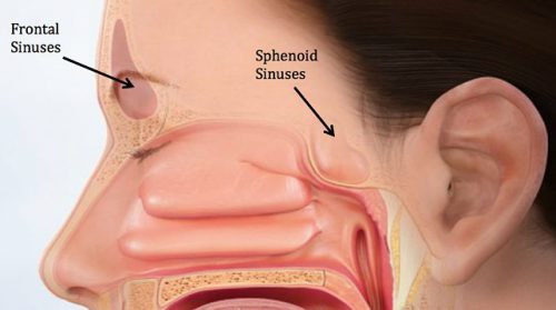
Here’s the normal anatomy:

sinus-fracture-treatment


Source: www.facialtraumamd.com

There are two “tables”, the anterior and the posterior. The anterior is covered with skin and a small amount of subcutaneous tissue. The posterior table is separated from the brain by the meninges.

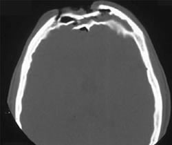
Here’s an image of an open fracture involving both tables. Note the underlying pneumocephalus.

frontal_sinus1

A third of injuries violate the anterior table, and two thirds violate both. Posterior table fractures are very rare. A third of all patients will develop a CSF leak, typically from their nose.

These fractures may be (rarely) identified on physical exam if deformity and flattening is noted over the forehead. Most of the time, these patients undergo imaging for brain injury and the fracture is found incidentally. Once identified, go back and specifically look for a CSF leak. Clear fluid in the nose is, by definition, CSF. Don’t waste time on a beta-2 transferring (see below).

If a laceration is clearly visible over the fracture, or if a CSF leak was identified, notify your maxillofacial specialist immediately. If more than a little pneumocephalus is present, let your neurosurgeon know. Otherwise, your consults can wait until the next morning.

In general, these patients frequently require surgery for the fracture, either to restore cosmetic contours or to avoid mucocele formation. However, these are seldom needed urgently unless the fracture is an open fracture with contamination or there is a significant CSF leak. If in doubt, though, consult your specialist.

Related posts:

Print Friendly, PDF & Email

Is Decompressive Craniectomy Any Better Than Craniotomy?

Severe TBI consists of a primary injury to the brain, followed by swelling, vascular, and ischemic problems which may cause a secondary injury. Much of the critical care management of this injury involves avoiding or ameliorating secondary injury. This is typically accomplished via medical means first, and through surgical procedures when medical management is insufficient.

Two types of surgical decompression are currently practiced: craniotomy and evacuation of blood/clot, and decompressive craniectomy with removal of a bone flap. The latter can be performed prophylactically before severe swelling occurs, or therapeutically as a damage control procedure when ICP is refractory to all other measures.

There has been a decades-old debate as to whether craniectomy, which is a major undertaking with months of skull/bone flap management, is actually worthwhile. Most studies have examined the utility of damage control craniectomy for refractory ICP. The results have not really been convincing one way or the other.

But what about prophylactic decompressive craniectomy (DC) to avoid future ICP problems while the patient is in the ICU? The surgical group at the University of Arizona at Tucson performed a five year retrospective review of their experience. Using propensity score matching, they identified 99 severe TBI patients who underwent DC (33) or craniotomy only (CO, 66). A power analysis showed that this sample size should be sufficient to demonstrate a significant difference.

Here are the factoids:

  • Both groups were similar with respect to age, GCS, ISS, AIS-head, and type of bleed
  • 26% died and 63% were discharged to rehab or skilled nursing facility
  • When comparing DC to CO groups, there were no differences in mortality, discharge to skilled nursing facility, discharge GCS or Glasgow Outcome Scale
  • There were more complications in the DC group, including shunt insertion for hydrocephalus (9% vs 0%), and reoperation (12% vs 2%)
  • Rates of wound infection and ventriculitis were the same for both groups (0-3%)

Bottom line: Although the study is small, it supposedly had enough patients for identification of significant differences. And basically, it didn’t show a positive difference for prophylactic decompressive craniectomy. There is certainly some opportunity for selection bias by the neurosurgeons that cannot be controlled for by this retrospective design. But it is yet another piece of the decompressive craniectomy puzzle. 

Overall, the literature support for either prophylactic or damage control craniectomy is not very strong. If it were, we would have identified some real benefits by now. What we don’t know is if there are specific subgroups of severe TBI patients who might benefit from it. So if your center is not involved in a project to study this, you should probably ask your neurosurgeons to base their practice only on what we know about this procedure to date. 

Print Friendly, PDF & Email