Category Archives: Head

What You Need To Know About: Frontal Sinus Fractures

Fracture of the frontal sinus is less common than other facial injuries, but can be more complex to deal with, both in the shorter and longer terms. These are generally high energy injuries, and facial impact in car crashes is the most common mechanism. Fists generally can’t cause the injury, but blunt objects like baseball bats can.

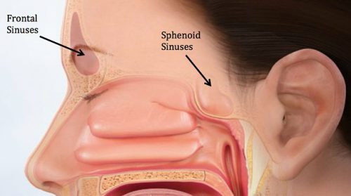
Here’s the normal anatomy:

sinus-fracture-treatment


Source: www.facialtraumamd.com

There are two “tables”, the anterior and the posterior. The anterior is covered with skin and a small amount of subcutaneous tissue. The posterior table is separated from the brain by the meninges.

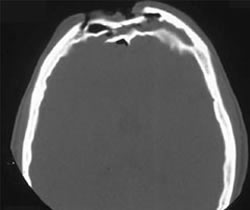
Here’s an image of an open fracture involving both tables. Note the underlying pneumocephalus.

frontal_sinus1

A third of injuries violate the anterior table, and two thirds violate both. Posterior table fractures are very rare. A third of all patients will develop a CSF leak, typically from their nose.

These fractures may be (rarely) identified on physical exam if deformity and flattening is noted over the forehead. Most of the time, these patients undergo imaging for brain injury and the fracture is found incidentally. Once identified, go back and specifically look for a CSF leak. Clear fluid in the nose is, by definition, CSF. Don’t waste time on a beta-2 transferring (see below).

If a laceration is clearly visible over the fracture, or if a CSF leak was identified, notify your maxillofacial specialist immediately. If more than a little pneumocephalus is present, let your neurosurgeon know. Otherwise, your consults can wait until the next morning.

In general, these patients frequently require surgery for the fracture, either to restore cosmetic contours or to avoid mucocele formation. However, these are seldom needed urgently unless the fracture is an open fracture with contamination or there is a significant CSF leak. If in doubt, though, consult your specialist.

Related posts:

Antihypertensive Treatment In Acute TBI

Yes, we know high blood pressure can be bad. Over the long term, it can accelerate atherosclerotic heart disease and pound away at the kidneys and brain. And when it is acutely elevated to critical levels, it can lead to stroke.

But is it always bad in trauma? Trauma hurts like hell, so it’s no wonder than many of our patients (not suffering blood loss of course) are hypertensive.  But how often have you seen this scenario occur:

An elderly patient fell from standing, striking her head. She is brought to your ED by ground EMS. She has a GCS of 8 (E1 V3 M4) with a BP of 200/130 and pulse of 56.  This meets your trauma activation criteria and the team assembles to meet the patient.

As you move her onto the bed, one of your colleagues calls out for some nicardipine to control the pressure. Is this a wise move? Remember the First Law of Trauma:

Any anomaly in your trauma patient is due to trauma, no matter how unlikely it may seem.

What else can cause hypertension and bradycardia in your trauma patient? In this case, certainly a subdural or epidural hematoma.

And why is that happening? Because the intracranial pressure is elevated from the space-occupying lesion. Remember the formula for cerebral perfusion pressure (CPP):

CPP = MAP – ICP

Where MAP = mean arterial pressure and ICP = intracranial pressure.  Normally the MAP is around 90 torr and ICP is about 10 torr. Thus, the normal CPP is approximately 80. The range is 60  to well over 100 torr, and flow autoregulation keeps brain perfusion constant over this range.

But let’s say that we are psychic and know the ICP of our patient to be 60 because of a large subdural hematoma. Her current CPP is 150 – 60 or about 90 torr. What happens if we start her on a nicardipine drip or some other antihypertensive medication? We can certainly normalize the blood pressure to 120/80. But now her CPP drops to 90  – 60 = 30 torr!

Congratulations, you have just shut down circulation to her brain!

Bottom line: Think first before calling for antihypertensive medications in patients who may have increased intracranial pressure. You may be sabotaging the only mechanism protecting their brain while you are calling your neurosurgeon for help. Your top priority is to get them to the CT scanner while permitting that pressure. If it turns out that there is no evidence for pathology that would lead to increased ICP, then turn to the antihypertensive agents to help protect against stroke. 

Is Intubation For Low GCS Necessary? Dangerous?

More dogma? I was taught that as the Glasgow Coma Scale (GCS) score drops toward 8, the higher the consideration of intubating the patient. And that a GCS 8 was pretty much an absolute indication for inserting the endotracheal tube. The rationale was that the more obtunded the patient was, the less able they were to protect their airway.

Even ATLS, our trauma textbooks, and practice guidelines from the likes of EAST recommend intubation for GCS 8 and less.

Having said that, I know many of you have been in a situation where you have a patient with GCS 8 or so, and they are lying there breathing peacefully with good oxygenation and ventilation. Do you really need to put in that tube? And we also tend to be very forgiving with obtunded children, avoiding premature intubation there as well.

Intubation is not a benign procedure. There is the potential for mayhem during the process, ICU admission will be required, and a host of ventilator and sedation-related complications are possible once the patient arrives there.

The trauma group at LAC + USC decided to look into this. They performed a five-year retrospective study of data from the TQIP database. A subset of patients was selected with isolated blunt head injury and GCS 7-8 who did not require immediate operation upon arrival. They were divided into intubation and non-intubation groups, and these were further subdivided into intubation within an hour of arrival, intubation after an hour, and never intubated groups.

Here are the factoids:

  • A total of 2,727 patients were studied; about two thirds were intubated within an hour, a quarter were never intubated, and the remaining 9% were intubated after the first hour
  • Immediately intubated patients were significantly younger and had fewer comorbidities
  • Mortality was 19% in the immediate intubation group vs 27% in the delayed group vs 11% in the never intubated group
  • Complications were significantly higher after immediate intubation, particularly DVT and ventilator associated pneumonia (VAP)
  • Regression analysis indicated that immediate intubation was independently associated with mortality compared to late or never intubated patients
  • Using additional regression testing, the authors concluded that the following two subsets of patients would benefit most from early intubation:
    • Younger patients (age < 45) with head AIS 5
    • Patients age <65 with head AIS 5

The authors recommend that “future research focus on more adequate parameters to identify patients requiring immediate intubation and should avoid an isolated fixed GCS threshold.”

Bottom line: This is a difficult paper to understand (at least for me). It looks like the authors are saying we should avoid immediate intubation of severe TBI patients with depressed GCS to reduce mortality and complications.

But you need to read the whole paper closely to really get it. First, let’s look at those mortality figures. The mortality in the three groups was:

  • intubated < 1 hour after arrival – 18.7% (from n = 1,866)
  • intubated > 1 hour after arrival – 27.4% (from n = 223)
  • never intubated – 11.4% (from n = 638)
  • If you combine the last two lines you get the mortality in the non-immediate intubations = 15.5% (from n = 861)

The authors then claim that the mortality for immediate intubation is greater than non-immediate intubation (the other two groups). This may be somewhat misleading, because the delayed intubation group actually has a higher mortality than the immediate group (27%)! This fact is covered up by combining delayed intubation with the never intubated group, bringing the number down to 15.5%!! Why shouldn’t you say that intubating the patient at any time is bad, immediate or delayed??

They suggest some criteria to try to focus in on the patients who really need intubation: younger patients (age < 45 or < 65) with head AIS 5 and GCS 7. Unfortunately, you can’t determine which patients have an AIS 5 in their head without a head CT, which may push them into the higher mortality delayed intubation group.

Remember, this type of study can only show an association, not cause and effect. The authors suggest that early intubation results in more deaths and complications. My suspicion is that patients with severe TBI don’t do poorly because they were intubated. I believe that they were intubated because the clinicians feared that they would do poorly. Unfortunately, this is information that can only be gleaned from a prospective study, not a retrospective database review.  And no amount of statistical manipulation or regression analysis can make up for this shortcoming.

This is yet another one of those studies that ends by concluding that a better study should be done. That would be okay if this one actually provided a hint that the endeavor would be worthwhile. But it doesn’t. I didn’t really learn anything from it, unfortunately.

So I still heartily recommend using your existing training, guidelines, and judgement to intubate these patients early and safely!

Reference: Isolated traumatic brain injury: Routine intubation for GCS 7 or 8 may be harmful! J Trauma, publish ahead of print, DOI: 10.1097/TA.0000000000003123, Feb 16, 2021.

Best of EAST #8: Reversing Antithrombotic Drugs After Severe TBI

Falls are the most common mechanism of injury at a majority of trauma centers these days. And due to the escalating number of comorbidities in our older population, more and more are taking some kind of anticoagulant or antiplatelet medication. And as all trauma professionals know, falling down and failure to clot do not mix well.

A variety of reversal regimens have been developed, including Vitamin K, plasma or platelet infusion, prothrombin complex concentrate, andexxanet, or idarucizumab depending on the agent. But when it comes to evaluating the efficacy of these agents, there are two important questions that need to be answered:

  1. Does the regimen reverse or neutralize the offending agent?
    and more importantly
  2. Does the regimen have a positive effect, i.e. reduce mortality and/or complications?

This last question has been problematic, especially for the direct oral anticoagulant drugs (DOACs). They are very expensive, but there has been little, if any, evidence that they improve mortality.

A study from the University of Florida at Jacksonville, and sponsored by EAST was performed last year. It was a multi-center, prospective, observational study of data provided by 15 US trauma centers. They collected data on the agents used, reversal attempts, and comorbidities in injured patients taking these drugs, and analyzed for head injury severity and mortality.

Here are the factoids:

  • There were a total of 2913 patients in the study, 46% on aspirin (ASA), 13% taking ASA and a P2Y12 inhibitor (one of the -grels), 11% on warfarin, 4% on ASA + warfarin, 13.5% on a Factor Xa inhibitor, and 6% on a Xa inhibitor + ASA
  • Patients on platelet blockers (P2Y12 inhibitor) had the highest mean ISS at 9
  • Warfarin was associated with a higher abbreviated injury score (AIS) for head, 1.2
  • Controlling for ISS, comorbidities, ISS, and initial SBP, warfarin + ASA had the highest head ISS with an odds ratio of 2.1 (with the lower confidence interval value of 1.19)
  • Reversal of antiplatelet therapy with DDAVP was not successful, with no change in mortality (87% with reversal and 93% without)
  • Reversal of Xa inhibitors with plasma or PCC was also unsuccessful with a mortality of 100% with reversal and 95% without

The authors concluded that reversal attempts for antiplatelet therapy or Factor Xa inhibitors did not decrease mortality, and shared the observation that combination therapies posed the most risk for severity of head injury.

My comments: Remember, the first thing to do is look at the study group. The authors did not share the inclusion or exclusion criteria for the study in the abstract, so we are a little in the dark here.

The next item that makes this study difficult to interpret (and perform) is the fact that nearly a quarter are on combination therapy for their anticoagulation. So even though nearly 3,000 patients were studied, many of the medication subgroups had only a few hundred subjects. The aspirin group was the largest, with 1,338. This makes me wonder if the overall study had the statistical power to find subtle differences in their outcome measures and support the conclusions.

Now have a look at one of the results tables:

In reviewing the demographic data, the concept of statistical significance vs clinical significance quickly comes to mind. Somehow, age, ISS, head AIS, mortality, and SBP are significantly different between some of the groups. Yet if you examine the specific values across most of the rows, there is little difference (e.g SBP ranges from 137 to 147, ISS from 7-9, mortality from 2-7%). These are all clinically identical. The only row that means much to me is the top one telling how many patients are in a group.

Here are my questions for the authors and presenter:

  1. Tell us about the study design, especially the inclusion and exclusion criteria. Were there any? How might this have influenced the study group?
  2. Please comment on your perception of the statistical power of the study, especially with seven groups of patients, each with relatively small numbers.
  3. Do you have information on the variety of reversal agents used? Were there any standards? Could this have contributed to the mortality in some of the groups?
  4. Do you have any clinical recommendations based on your findings? If not, what is the next step in examining this group of patients?

My bottom line is that I’m not sure that this study has the power to show us any significant differences. And looking at the information table and logistic regression results (odds ratio confidence intervals close to 1), I’m not really able to learn anything new from it. I’m hoping to learn a lot from the live presentation!

Reference: EAST MCT: comparison of pre-injury antithrombotic use and reversal strategies among severe TBI patients. EAST 2021, Paper 19.

Everything You Wanted To Know About: Cranial Bone Flaps

Patients with severe TBI frequently undergo surgical procedures to remove clot or decompress the brain. Most of the time, they undergo a craniotomy, in which a bone flap is raised temporarily and then replaced at the end of the procedure.

But in decompressive surgery, the bone flap cannot be replaced because doing so may increase intracranial pressure. What to do with it?

There are four options:

  1. The piece of bone can buried in the subcutaneous tissue of the abdominal wall. The advantage is that it can’t get lost. Cosmetically, it looks odd, but so does having a bone flap missing from the side of your head. And this technique can’t be used as easily if the patient has had prior abdominal surgery.

2. Some centers have buried the flap in the subgaleal tissues of the scalp on the opposite side of the skull. The few papers on this technique demonstrated a low infection rate. The advantage is that only one surgical field is necessary at the time the flap is replaced. However, the cosmetic disadvantage before the flap is replaced is much more pronounced.

3. Most commonly, the flap is frozen and “banked” for later replacement. There are reports of some mineral loss from the flap after replacement, and occasional infection. And occasionally the entire piece is misplaced. Another disadvantage is that if the patient moves away or presents to another hospital for flap replacement, the logistics of transferring a frozen piece of bone are very challenging.

4. Some centers just throw the bone flap away. This necessitates replacing it with some other material like metal or plastic. This tends to be more complicated and expensive, since the replacement needs to be sculpted to fit the existing gap.

So which flap management technique is best? Unfortunately, we don’t know yet, and probably never will. Your neurosurgeons will have their favorite technique, and that will ultimately be the option of choice.

Reference: Bone flap management in neurosurgery. Rev Neuroscience 17(2):133-137, 2009.