Category Archives: Complications

Nausea In The Trauma Bay: Gastric Tube vs Anti-Emetic Drugs?

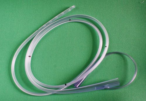
Nausea and vomiting are common problems in trauma patients, particularly those in a trauma activation. Inciting factors include pain, full stomach from food eaten before the event or blood swallowed after, or reaction to pain medications. For years, trauma professionals reached for the lowly gastric tube to evacuate stomach contents to “solve” the problem.

But how many of you have seen a patient forcefully empty their stomach as soon as the tube touches the oropharynx? And of course, your patient is lying supine, so the vomitus goes straight up, then back down into their airway. And if their mental status is not quite right, they may aspirate, causing even bigger problems.

We’ve had anti-emetic medications for a long time, some more effective than others. Only recently have we begun to rely on these as a first line defense in the trauma resuscitation room. But do they work? Are they safer?

The University Medical Center Utrecht in the Netherlands looked at this problem. They changed their policy from inserting a gastric tube to administering anti-emetics at the beginning of 2014. They studied their experience for the 6 months before and 6 months after the policy change. They inserted an orogastric (OG) tube preferentially before the switch, and used ondansetron and/or metoclopramide after.

Here are the factoids:

  • A total of 1446 trauma patients were admitted during this period. After excluding patients who were intubated or did not complain of nausea, 453 were analyzed (30%)
  • 20% of patients who had an OG tube placed vomited vs only 3% receiving medication (significant)
  • After therapy, 14% of patients receiving an OG were still nauseated vs only 2% getting meds (also significant)
  • 3 patients vomited and aspirated after OG placement, and 1 developed a pneumonia. 2 patients became bradycardic and med administration, and one developed QT-prolongation

Bottom line: This is a relatively small, retrospective study. Furthermore, the choice of gastric tube route (oral) is a setup for gagging and vomiting. Nasogastric tubes are a bit less noxious, but can’t be inserted in all patients (see tomorrow’s post). Even so, the use of anti-emetics in trauma patients complaining of nausea seems like the kinder, gentler way to go. 

Which drug to use? Previous studies have shown that ondansetron 4mg is as effective as 8mg, and that this drug is about equally as effective as metoclopramide. There is also some evidence that giving both is more effective than just giving one.

Gastric tubes are still important, particularly in the comatose patient. But since these patients are at risk for cribriform plate injury, only the oral route should be used.

Reference: Analysis of two treatment modalities for the prevention of vomiting after trauma: orogastric tube or anti-emetics. Injury (accepted manuscript, in press) online 8 July 2017.

Print Friendly, PDF & Email

Aspirin For DVT Prophylaxis In Trauma

The use of mechanical and pharmacologic prophylaxis for prevention of deep venous thrombosis (DVT) and venous thromboembolism (VTE) in trauma patients is nearly universal. However, no matter how closely we adhere to existing guidelines, some patients will develop these conditions. Indeed, about 80% of patient who suffer some type of VTE event were receiving prophylaxis at the time.

Trauma is a major factor in causing hypercoagulability. Although current chemoprophylaxis focuses on clotting factors, platelets play a big part in the clot formation process. Our usual drugs, though (various flavors of heparin), have no effect on them.

What about adding aspirin to the regimen? My orthopedic colleagues have been requesting this for years. There is a reasonable amount of data in their literature that it is effect in patients with knee arthroplasty only. As usual, it is misguided to try to generalize management based on experience from one specific body region or operation.

A single Level I trauma center reviewed its data on aspirin prophylaxis for trauma patients. They reviewed their registry data from 2006 to 2011. They identified 172 trauma patients with duplex ultrasound proven DVT. These patients were matched with 1,901 control patients who underwent at least one duplex and never developed DVT. Matching was performed carefully to ensure that age, probability of death, number of DVT risk factors, and presence of TBI were similar. The total number of matched patients studied was 110.

And here are the factoids:

  • About 7% of patients with DVT were on aspirin at the time of their injury, vs 14% of the matched controls
  • 7% were taking warfarin, and 4% were taking clopidogrel
  • Analysis showed that patients taking aspirin had a significantly decreased chance of DVT after injury
  • On further analysis, it was found that this effect was only significant if some form of heparin was given for prophylaxis as well.

Bottom line: So before you run off and start giving your patients aspirin, think about what this study really said. Patients taking aspirin before their injury and coupled with heparin after their injury have a lower rate of DVT. It gives us no guidance as to whether adding aspirin after the fact, or using aspirin alone, are useful.  And we still don’t know if any of this decreases pulmonary embolism or mortality rates.

Related posts:

Reference: Aspirin as added prophylaxis for deep vein thrombosis in trauma: a retrospective case-control study. J Trauma 80(4):625-30, 2016.

Print Friendly, PDF & Email

Prevnar 13 And Spleen Trauma

Recently, I’ve noticed television commercials for Prevnar-13, a 13-valent pneumococcal vaccine for immunocompromised or asplenic adults. And interestingly, I noticed that the CDC has now added a recommendation such that these patients receive this vaccination, and then the good old 23-valent vaccine (Pneumovax) 8 weeks later.

WTF? Patients with splenectomy (or significant angio-embolization) for trauma are considered functionally asplenic. And although the data for immunization in this group is weak, giving triple vaccinations with pneumcoccal, H. flu, and meningococcal vaccines has become a standard of care.

This was difficult enough already because there was debate around the best time to administer: during the hospital stay or several weeks later after the immune system depression from trauma had resolved. The unfortunate truth is that many trauma patients never come back for followup, and so don’t get any vaccines if they are not given during the hospital stay.

And then came the recommendation a few years ago to give a 5-year booster for the pneumococcal vaccine. I have a hard time remembering when my last tetanus vaccine was to schedule my own booster. How can I expect my trauma patients to remember and come back for their pneumococcal vaccine booster?

So what do we do with the CDC Prevnar-13 recommendation? If we add it, it means that we give Prevnar while the patient is in the hospital, and then hope they come back 8 weeks later for their Pneumovax. And then 5 years later for the booster dose. Huh?

Looking at the package insert, I read that Pneumovax protects against 23 serotypes of S. Pneumo, which represent 85% of most commonly encountered strains out there. So it’s not perfect. Prevnar-13 protects against 13 serotypes, and there is no indication as to what percent of strains encountered are protected against.

So I decided to dig deeper and look at the serotypes included in each vaccine. Here they are:

  • Pneumovax: 1, 2, 3, 4, 5, 6B, 7F, 8, 9N, 9V, 10A, 11A, 12F, 14, 15B, 17F, 18C, 19F, 19A, 20, 22F, 23F, and 33F
  • Prevnar: 1, 3, 4, 5, 6A, 6B, 7F, 9V, 14, 18C, 19A, 19F, and 23F

I bolded the serotypes in Prevnar-13 not found in the Pneumovax vaccine. There was only one, serotype 6A. Unfortunately, it’s nearly impossible to find the prevalence by serotype, and it varies geographically and over time.

Bottom line: I’m not an epidemiologist. But making a set of vaccination rules more complicated for a complex population, and for indications that are a bit weak in the first place, seems unwise. Especially since the added vaccine offers protection for only one more serotype of Pneumococcus.

So please help me out here. Show me something I’m missing. Otherwise, I’ll stick to the original three vaccines, and try to remind my patients to get that booster five years down the road.

Related posts:

Reference: Use of 13-Valent Pneumococcal Conjugate Vaccine and 23-Valent Pneumococcal Polysaccharide Vaccine for Adults with Immunocompromising Conditions: Recommendations of the Advisory Committee on Immunization Practices (ACIP). MMWR 61(40):816-819, October 12, 2012.

Print Friendly, PDF & Email

Incidental Finding: Gas In The Spleen After Embolization

Most solid organ injury practice guidelines include angioembolization in part of the pathway. But very few require re-imaging at any point to see how the liver or spleen are coming along.

But every once in a while another condition arises, or symptoms worsen unexpectedly, causing us to get another CT scan that includes the abdomen and pelvis. And sometimes we see things that we wouldn’t normally see, like air bubbles in the organ that was embolized.

So what is okay, and what requires some kind of intervention? Our friends at ShockTrauma in Baltimore looked at this in 2001 and can provide some pretty good guidance. They reviewed patients who underwent CT scan both before and after embolization over about 2.5 years. They performed the post-embolization scans for specific indications like fevers, elevated WBC count (!), increasing abdominal pain, or an episode of hypotension. A total of 53 patients were studied.

Here are the factoids:

  • 24 patients underwent embolization of the main splenic artery, 22 had selective embolization of part of the spleen, and 7 had both
  • Splenic infarcts occurred in 63% of patients with main artery embolization, but were large (> 50% of the parenchyma) in only 20% of those
  • Infarcts occurred in 100% of selective embolizations, but were small (< 50%) in 93% of cases
  • Infarcts occurred in 71% of patients with both main and selective embolization, and most were small (80%)
  • Seven (13%) patients developed gas bubbles in the spleen, and was usually present for 1-7 days before disappearing
  • One patient developed increasing gas with pneumoperitoneum and underwent splenectomy for a splenectomy for abscess

This picture that shows tiny bubbles in the spleen parenchyma that represent “normal” gas after embolization:

And the following one shows an air/fluid collection in the spleen that indicates an abscess:

Bottom line: Tiny bubbles in the spleen (and probably the liver) occur normally after angioembolization. They usually develop within an area of infarction, and most are benign. It is possible for them to evolve into a splenic abscess, but unlikely. Many embolization patients develop fevers at some point, and most have an elevated WBC count. So in most cases, you can ignore this incidental finding, as long as your patient has mild symptoms.

However, if the patient develops high fevers, very elevated WBC (> 25K), increasing abdominal or flank pain, and the spleen develops an air/fluid level, an abscess is forming. Despite what your radiologist might suggest, catheter drainage is not a good idea. The tubes are too small to remove the slurry that is generally found within the abscess. A trip to the OR is the only effective treatment, and splenectomy is generally the only option.

Related posts:

Reference: CT Findings after Embolization for Blunt Splenic Trauma. J Vasc Intervent Radiol 12(2):209-214, 2001.

Print Friendly, PDF & Email

Retained Hemothorax Part 3: VATS

I’ve written about the use of lytics to treat retained hemothorax over the past few days. Although it sounds like a good idea, we just don’t know that it works very well. And they certainly don’t work fast. Lengths of stay were on the order of two weeks in both studies reviewed.

The alternative is video assisted thoracoscopic surgery (VATS). So let’s take a look at what we know about it. This procedure is basically laparoscopy of the chest. A camera is inserted, and other ports are added to allow insertion of instruments to suck, peel, and scrape out the hemothorax.

A prospective, multi-center study was performed over a 2 year period starting in 2009. Twenty centers participated, contributing data on 328 patients with retained hemothorax. This was defined as CT confirmation of retained blood and clot after chest tube placement, with evidence of pleural thickening.

Here are the factoids:

  • 41% of patients had antibiotics given for chest tube placement (this is interesting given the lack of consensus regarding their effectiveness!)
  • A third of patients were initially managed with observation, and most of them (82%) did not need any further procedures (83 of 101 patients)
  • Observation was more successful in patients who were older, had smaller hemothoraces (<300cc), smaller chest tubes (!!, <34 Fr), blunt trauma, and peri-procedure antibiotics (?)
  • An additional chest tube was inserted in 19% of patients, image guided drain placement in 5%, and lytics in 5%. Half to two-thirds of these patients required additional management.
  • VATS was used in 34% of patients. One third of them required additional management including another chest tube, another VATS, or even thoracotomy.
  • Thoracotomy was most likely required if there was a diaphragm injury or large hemothorax (<900cc)
  • Empyema and pneumonia were common (27% and 20%, respectively)

Bottom line: There’s a lot of data in this paper. Most notably, many patients resolve their hemothorax without any additional management. But if they don’t, additional tubes, guided drain placement, and lytics work only a third of the time and contribute to additional time in the hospital. Even VATS and thoracotomy require additional maneuvers 20-30% of the time. And infectious complications are common. This is a tough problem!

Tomorrow, I’ll try to roll it all together and suggest an algorithm to try to optimize both outcomes and cost.

Posts in this series:

Reference: Management of post-traumatic retained hemothorax: A prospective, observational, multicenter AAST study. 72(1):11-24, 2012.

Print Friendly, PDF & Email