Category Archives: Complications

The Final X-Ray In Damage Control Surgery

Damage control surgery for trauma is over 20 years old, yet we continue to find ways to refine it and make it better. Many lives have been saved over the years, but we’ve also discovered new questions. How soon should the patient go back for definitive closure? What is the optimal closure technique? What if it still won’t close?

One other troublesome issue surfaced as well. We discovered that it is entirely possible to leave things behind. Retained foreign bodies are the bane of any surgeon, and many, many systems are in place to avoid them. However, many of these processes are not possible in emergent trauma surgery. Preop instrument counts cannot be done. Handfuls of uncounted sponges may be packed into the wound.

I was only able to find one paper describing how often things are left behind in damage control surgery (see reference below), and it was uncommon in this single center study (3 cases out of about 2500 patients). However, it can be catastrophic, causing sepsis, physical damage to adjacent organs, and the risk of performing an additional operation in a sick trauma patient.

So what can we do to reduce the risk, hopefully to zero? Here are my  recommendations:

  • For busy centers that do frequent laparotomy or thoracotomy for trauma and have packs open and ready, pre-count all instruments and document it
  • Pre-count a set number of laparotomy pads into the packs
  • Use only items that are radiopaque or have a marker embedded in them. This includes surgical towels, too!
  • Implement a damage control closure x-ray policy. When the patient returns to OR and the surgeons are ready to begin the final closure, obtain an x-ray of the entire area that was operated upon. This must be performed and read before the closure is complete so that any identified retained objects can be removed.

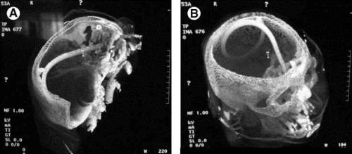
Tomorrow, a sample damage control closure x-ray.

Related post:

Reference: Retained foreign bodies after emergent trauma surgery: incidence after 2526 cavitary explorations. Am Surg 73(10):1031-1034, 2007.

Print Friendly, PDF & Email

Natural History of the Splenic Blush

In my last post, I described the two types of solid organ “blushes.” I also described my thoughts on the natural history of these findings. Now, a multicenter study on the natural progression of the splenic “blush” has just been published. I found this paper very interesting, because it challenged some of my own existing beliefs. But once I read it, my enthusiasm faded.

The Western Trauma Association sponsored a multicenter (17 Level I and II centers) review of data collected prospectively over an unspecified period of time. Patients were excluded if their injury was older than 24 hours, if they had a previous splenic injury, and if they had any number of diseases or hereditary conditions that might affect the spleen. Strict definitions of nonbleeding and actively bleeding injuries were applied, and detailed information on intervention and outcomes was collected.

Here are the factoids:

  • 200 patients were enrolled from 17 centers, but the paper does not state how long that took
  • 20% were low grade (1 or 2) and 80 % high grade (3-5)
  • 29% had a pseudoaneurysm, and 83% showed extravasation, which means that several patients had both
  • 15% underwent early splenectomy, 59% underwent angiography, and 26% were observed
  • For those with initial angiography, 6% had repeat angio and 7% eventually underwent splenectomy
  • Of those were were initially observed, 9% had delayed angio and 8% underwent splenectomy
  • Based on a read by an expert radiologist, an actively bleeding injury was associated with a 41% splenectomy rate
  • The authors conclude that the majority of patients with spleen injury with pseudoaneurysm or extravasation are managed with angio and embolization and that splenectomy remains a rare event (??)

Bottom line: This paper just doesn’t do it for me. The biggest problem is that it is what I call a “we do it the way we do it” study. It examines how 17 different centers evaluate and treat patients with significant splenic injury. There was no guidance or guideline on how to treat, so they each did it their way. And the number of patients was small.

They don’t tell us anything about the use or effectiveness of angio by grade. Or whether the specific hospitals routinely rely on angio rather than just going to the OR for high grade injuries (typically if angio response times are long).

Unfortunately, this paper gives the appearance of containing a lot of interesting stuff. But a 15% initial splenectomy rate is not a “rare event” in my book. Everything published here is at odds with what I’ve observed over the years for centers with well developed management guidelines and easy access to angio (< 5% splenectomy rate in hemodynamically stable patients with nonoperative management).

My recommendation is to send all stable patients with pseudoaneursym and/or extravasation to angio immediately! Yes, some will have nothing found by the time they get to angio, and you’ll have to come up with a plan at that point. But most have something wrong, and it won’t stop until it’s been plugged up (or your patient bleeds to death, whichever comes first)!

This article has all the right buzzwords: multicenter, prospective data, etc. But it’s already been moved to my recycle bin. 

Related post:

Reference: Natural history of splenic vascular abnormalities after blunt injury: A Western Trauma Association multicenter trial. J Trauma 83(6):999-1005, 2017.

Print Friendly, PDF & Email

Syncope Workup in Trauma Patients – Updated With CPG

Syncope accounts for 1-2% of all ED visits, and is a factor in some patients with blunt trauma, especially the elderly. If syncope is suspected, a “syncope workup” is frequently ordered. Just what this consists of is poorly defined. Even less understood is how useful the syncope workup really is.

Researchers at Yale retrospectively looked at their experience doing syncope workups in trauma patients. They were interested in seeing what was typically ordered, if it was clinically useful, and if it impacted length of stay.

A total of 14% of trauma patients had syncope as a possible contributor to their injury. The investigators found that the following tests were typically ordered in these patients:

  • Carotid ultrasound (96%)
  • 2D Echo (96%)
  • Cardiac enzymes (81%)
  • Cardiology consult (23%)
  • Neurology consult (11%)
  • EEG (7%)
  • MRI (6%)

Most of this testing was normal. About 3% of cardiac enzymes were abnormal, as were 5% of carotid imaging and 4% of echocardiograms.

Important! Of the patients who underwent an intervention after workup, 69% could have been identified based on history, physical exam, or EKG and did not depend on any of the other diagnostic tests.

Is it possible to determine a subset of this population that may show a higher yield for this screening? Surgeons at Temple University in Philadelphia found that there was little utility in using carotid duplex studies. They did note that patients with a history of heart disease were more likely to have an abnormal EKG, and that an abnormal EKG predicted an abnormal echo. Overall, only patients with a history of significant cardiac comorbidity, older age, and higher ISS had findings requiring intervention.

Bottom line: Don’t just reflexively order a syncope workup when there is a question of this problem. Think about it first, because the majority of these studies are nonproductive. They are not needed routinely in trauma patients with “syncope” as a contributing factor.  Obtain a good cardiac history, and if indicated, order an EKG and go from there. See the practice guideline proposed by the Temple group below. And be sure to include the patients primary doctor in the loop!

References:

  1. Routine or protocol evaluation of trauma patients with suspected syncope is unnecessary. J Trauma 70(2):428-432, 2011.
  2. Syncope workup: Greater yield in select trauma population. Intl J Surg, accepted for publication June 27, 2017.

Print Friendly, PDF & Email

The ICU Bounce Back

We’ve all experienced it. A seriously injured trauma patient is admitted to the ICU and begins the process of recovery. Everything looks well, and after a few days they’re transferred to a ward bed. But then they languish, never really doing what we expect. Finally (and usually in the middle of the night), they begin to look bad enough where we have to transfer them back to the ICU. Before or after the call to the Rapid Response Team. Yes, it’s the feared “unexpected readmission to ICU.”

What’s the problem here? A failure of the ICU team? Did they send the patient out too soon? Did we all miss something about the patient? And is there any way we can avoid this problem? The major issue is that these “bounce backs” tend to do poorly compared to patients who successfully stay in their ward bed. Estimates are that mortality for patients successfully and finally discharged from the ICU range from 4-8%, whereas the mortality in bounce back patients is 20-40%!

Researchers at the Medical University of South Carolina in Charleston looked at the characteristics that defined the bounce back patient. They reviewed nearly 2000 patients discharged from their trauma ICU and analyzed the variables that predicted an unplanned bounce back. They noted the following interesting factoids:

  • More than two thirds of bounce backs occurred within 3 days
  • Males, patients with an initial GCS < 9, transfer during the day shift  were the major risk factors
  • More comorbidities was associated with a higher chance of bounce back
  • Mortality in the bounce back group was 20%
  • The most common immediate factors causing bounce back were respiratory failure or bleeding

Bottom line: This is an intriguing single-institution study that supports my own personal observations. Fewer bounce backs occur at night because staffing tends to be lower and there is more resistance to transfers out of the ICU then. Both the ICU team and the ward team need to scrutinize every transfer carefully. Significant head injury or the presence of medical comorbidities should trigger a careful assessment to make sure that the transfer is appropriate. Otherwise, your patient may be placed in unnecessary jeopardy.

Next, I’ll discuss when an unexpected return to ICU is not an unexpected return!

Reference: Intensive care unit bounce back in trauma patients: An analysis of unplanned returns to the intensive care unit. J Trauma 74(6):1528-1533, 2013.

Print Friendly, PDF & Email

Top 10 Worst Complications: #1 Nasocerebral Tube

Minor complications from nasogastric tube insertion occur relatively frequently. Emesis is fairly common when the gag reflex is stimulated by the tube in the back of the oropharynx. An infrequent but possibly fatal one is insertion through the cribriform plate. 

The cribriform plate is located directly posterior to the nares and is part of the ethmoid bone. It is very porous in nature and weaker than the surrounding portions of the ethmoid. It is easily fractured, and can be seen is association with basilar skull fractures. This is one source for rhinorrhea in patients with these fractures.

Cribriform fracture is a contraindication to unprotected insertion of a nasogastric tube. If you look at the sagittal section below, the plate lies directly behind the nares. When inserting the NG tube, we are usually taught to aim the tube straight back. Unfortunately, this aims it directly at the cribriform. If a fracture is present, it is possible that you may be inserting a nasocerebral tube!

Cribriform plate - sagittal section

The usual symptoms when this occurs consist of immediate neurologic deterioration to coma, and a unilateral or bilateral blown pupil. The tube must not be withdrawn, because it will cause significant injury to the base of the brain. A stat neurosurgical consultation must be obtained, and if the patient is salvageable, the tube must be withdrawn through a craniectomy.

To avoid this dreaded complication, identify patients at risk for cribriform injury. They are:

  • patients with signs of trauma from eyebrows to zygoma
  • comatose patients
  • patients with signs of basilar skull fracture (Battle’s sign, raccoon eyes, oto- or rhinorrhea)

If your patient is at risk, follow these guidelines:

  • first, does the patient really need a gastric tube?
  • if comatose, insert an orogastric tube
  • if awake, don’t put the tube in their mouth, as they will gag continuously. Instead, place a lubricated, curved nasal airway. Then lube up a slightly smaller Salem sump tube and pass it through the airway.
Print Friendly, PDF & Email