Category Archives: Complications

What Does A Retained Surgical Sponge Look Like?

Surgeons and surgical residents rarely see these. And because it’s so uncommon, they frequently don’t recognize the telltale findings on radiographic studies. The TSA runs into the same problem in screening passengers for weapons and other hazards at airports. But it’s the bane of any surgeon’s existence. And it’s a major reason why OR personnel take such great pains to account for everything in the room. It is a catastrophe, and always a preventable one, when some piece of equipment goes missing and ends up left inside a patient.

A number of methods have been developed to try to eliminate this problem. They include careful counts, having someone record anytime anything is placed inside, x-rays, and most recently, RFID tags.

After counting, x-ray is the most common way to try to find missing objects. One would think that these foreign bodies would be easy to see. Metallic instruments are rather easy to spot. But many trauma professionals, even those who work in the OR, have never seen what a positive image of a sponge actually looks like. So here they are. You should never miss one on an xray now.

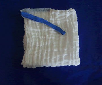
Surgeons typically use two types of sponges in the OR: Ray-Tec sponges and standard lap pads. Ray-Tecs look like a 4×8 piece of gauze with a mysterious blue string woven throughout it. The string is the only part that shows up on x-ray, and it is very thin and somewhat hard to see. Here are some Ray-Tec sponges outside the body:

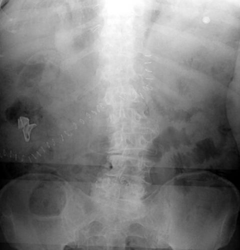
And here’s one that was left inside. Note the little squiggle in the left lower quadrant and how easy it is to overlook.

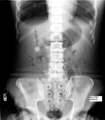
On the other hand, a laparotomy pad is a 4×4 folded cloth pad that unfolds into a larger pad. It has a blue radiopaque tag sewn in the corner, extending along one edge of the pad. Here’s what they look like outside the body:

And here’s one inside a patient. Note the irregular object in the right upper quadrant. Many times the tag is scrunched up and doesn’t look like one.

Bottom line: It’s important for anyone who works in the OR on any body part to be familiar with the appearance of these tags on x-rays. Since it’s generally impossible to get accurate counts before or after a trauma procedure, always image the involved body cavity looking for these telltale signs before closing the patient.

Note: These images  were taken from the internet. Patients were not treated at Regions Hospital.

Print Friendly, PDF & Email

Consequences Of Embolizing Renal Injuries

In my last post, I noted that nonoperative management is the norm for dealing with high grade renal injuries. One of the possible options, angioembolization, was relatively infrequently used at only 6% of the time.

For management of other organs like the spleen, there are several angioembolization options. Depending on the type and severity of injury, selective (partial) or nonselective (main splenic artery) embolization can be carried out. For the liver, only selective embolization can be used. But what about the kidney? 

Are there consequences of nonselective renal embolization? Or should we always strive for selective control? The urology group of the University of Tennessee – Knoxville published a series of papers on their experience using embolization in patients with the most severe injuries (Grade 5). They retrospectively examined just over 3 years of admissions with this injury. Numbers were very small (6 men, 3 women).

But they also published a second paper, extending the review dates to capture one more male patient. And they followed this group for 1.5 to 5 years (mean 2.5 years) to determine if any delayed complications surfaced.

Here are the factoids:

  • Seven patients underwent full, nonselective embolization, and the other three had “super selective” embolization
  • All patients had control of bleeding without surgical intervention
  • Followup CT imaging showed no persistent extravasation or expanding hematoma
  • No patient developed complications, such as a retroperitoneal abscess, prolonged fever, or hypertension while in the hospital or during short-term followup
  • Most patients showed a very small increase in serum creatinine (mean 0.04), but one patient increased from 1.1 to 1.7
  • On longer term followup, one patient, age 51, developed hypertension 10 months after his injury. It is not possible to determine whether he was one of the 20% of older adults who develop hypertension, or whether it was due to the procedure. it was well-controlled with a single antihypertensive med.
  • None developed altered renal function, stones, chronic pain, fistula, or pseudoaneurysm

Bottom line: Obviously, the data is very limited with only 10 patients. However, it is very interesting to note that the majority of these patients underwent nonselective embolization of the renal artery without any adverse event. The one case of hypertension occurred with nonselective embolization, although I have seen several case reports where this occurs with selective embolization as well.

It is now well-accepted that high-grade renal injury can and should be managed nonoperatively if the patient’s hemodynamic status is reasonable. I recommend a trip to interventional radiology if the patient has active extravasation or a high-grade (Grade 4 or 5) injury, as these patients are at risk for loss of the entire kidney otherwise. Selective embolization can be attempted first, but don’t be shy to take out the entire organ if need be. 

References: 

  • Percutaneous embolization for the management of Grade 5 renal trauma in hemodynamically unstable patients: initial experience. J Urology 181:1737-1741, 2008.
  • Intermediate-term follow-up of patients treated with percutaneous embolization for Grade 5 blunt renal trauma. J Trauma 69(2):468-470, 2010.
Print Friendly, PDF & Email

Complications Of Iliac Artery Embolization

The main cause of mortality in patients with severe pelvic fractures is major hemorrhage. Over the years, trauma professionals have developed and tested a number of maneuvers to reduce mortality in these patients. These include wrapping or fixing the pelvis, embolization, and more recently, pre-peritoneal packing and REBOA.

Pelvic wrap/fixation and embolization have been around for a long, long time. For both, it’s been long enough so that we should have a fairly decent appreciation of the complications. For pelvic binders, they principally involve the skin. But aside for the potential access site complications (bleeding, pseudoaneurysm), angiography has been thought to be relatively benign.

But as with any medical procedure, especially invasive ones, there are risks. A paper published five years ago retrospectively reviewed the 13 year experience with pelvic angiography at UC Davis. Study patients were matched with controls who underwent angiography for pelvic fracture but not embolization. Short-term (within 30 days) and long-term complications were assessed while in hospital and by telephone survey. Mean followup time was 18 months.

Here are the factoids:

  • There were no differences in complications attributable to embolization within 30 days of the procedure
  • There were 5 cases of short-term skin sloughing or necrosis in 55 patients, and 4 of 5 occurred in patients with nonselective embolization. However, this was not a statistically significant complication.
  • Long-term complications such as buttock claudication or skin ulceration, pain, and impotence were not significantly different in embolized vs non-embolized patients
  • There was a significantly increased incidence of buttock, perineal, or thigh paresthesias in the long-term

Bottom line: Angiography with embolization is a very valuable tool in the management of complication pelvic fractures. Remember that a number of complications have been described:

  • Skin sloughing or necrosis
  • Buttock claudication, pain, paresthesias
  • Skin ulceration
  • Impotence

Other than an increase in paresthesias in the long-term, there did not appear that there was any difference in patients undergoing angiography with and without embolization. Although the numbers were small (100 patients total), this is the best study we have to date. Just keep in mind that complications are possible, and question your patients about them when they present for their followup visits.

Reference: Evaluation of Short-term and Long-term Complications after Emergent Internal Iliac Artery Embolization in Patients with Pelvic Trauma. J Vascular Interventional Rad 19(6):840-847, 2008.

Print Friendly, PDF & Email

Keeping Patients Warm In Your Trauma Bay

Hypothermia is the enemy of all trauma patients. It takes their potential bleeding problems and makes them exponentially worse. From the time you strip off their clothes in the trauma resuscitation room, they begin to cool down. And if you live in Minnesota like me (or some similar fun place), they start chilling even before that.

What can you do in the trauma bay to help avoid this potential complication? Here are some of the possibilities, and what I think of them. And I’ll also provide a practical tip to help keep your patient warm  while you can still do a full exam.

Outside

– Warming lights in the ambulance unloading area. I know lots of people look at this area and recommend them. Unfortunately, they don’t do a lot. Consider that your patient will move through this space quickly. While it may be cold, they’ll only spend a minute or so getting to the back door to the ED.

– How about the path from the helipad? If this is mostly outside, it can be a problem. If it’s wide open, there aren’t really a lot of options. Cover and heat it? Lots of $$$. Typically, flight crews working in winter climates have bundled up their patient very well, and this is the patient’s primary source of protection from the elements. If the pad is far away from the ED, consider a fancy golf cart to move them quickly, and perhaps get an even fancier one that has a heated enclosure.

Inside

– Heat the room! This only works on a moment’s notice if you have a smaller room or a really good heating system. Otherwise, you must keep it cranked it up at all times.

– Close the door! You will not be able to keep the room toasty unless you make sure the door is closed as much as possible. No doors? Then consider the next tips.

– Use radiant heating systems. Some EDs have lights in the ceiling, others have portable units that can be rolled over to your patient.

– Use hot fluids, especially in the winter. At a minimum, all blood products must be administered through a warmer, since they are only a few degrees above freezing. If it’s winter outside, or your patient is already cool, give all IV fluids through the warmer, too.

– Cover your patient. Keep a blanket warmer nearby, and pull several out at the beginning of each resuscitation.

– What about those fancy air blankets? Unfortunately, they are unwieldy. They’re all one piece, they try to fall of the patient all the time, and they limit access for your exam. But there is a solution!

Here’s a clever way to deal with this problem. Use my two-blanket trick. Don’t use just one warm sheet or blanket. Use two! Fold each one in half, so they are each half-length. Place one on the top half of the patient, the other at the bottom, overlapping slightly at the waist. Your whole patient is now covered and toasty. If you need to look at an extremity, fold the blanket that covers it over from right to left (or left to right) to uncover just the area of interest. To insert a urinary catheter, just open the area at the waist, moving the top sheet up a little, the bottom down a little. Voila!

Print Friendly, PDF & Email

The Final X-Ray In Damage Control Surgery

Damage control surgery for trauma is over 20 years old, yet we continue to find ways to refine it and make it better. Many lives have been saved over the years, but we’ve also discovered new questions. How soon should the patient go back for definitive closure? What is the optimal closure technique? What if it still won’t close?

One other troublesome issue surfaced as well. We discovered that it is entirely possible to leave things behind. Retained foreign bodies are the bane of any surgeon, and many, many systems are in place to avoid them. However, many of these processes are not possible in emergent trauma surgery. Preop instrument counts cannot be done. Handfuls of uncounted sponges may be packed into the wound.

I was only able to find one paper describing how often things are left behind in damage control surgery (see reference below), and it was uncommon in this single center study (3 cases out of about 2500 patients). However, it can be catastrophic, causing sepsis, physical damage to adjacent organs, and the risk of performing an additional operation in a sick trauma patient.

So what can we do to reduce the risk, hopefully to zero? Here are my  recommendations:

  • For busy centers that do frequent laparotomy or thoracotomy for trauma and have packs open and ready, pre-count all instruments and document it
  • Pre-count a set number of laparotomy pads into the packs
  • Use only items that are radiopaque or have a marker embedded in them. This includes surgical towels, too!
  • Implement a damage control closure x-ray policy. When the patient returns to OR and the surgeons are ready to begin the final closure, obtain an x-ray of the entire area that was operated upon. This must be performed and read before the closure is complete so that any identified retained objects can be removed.

Tomorrow, a sample damage control closure x-ray.

Related post:

Reference: Retained foreign bodies after emergent trauma surgery: incidence after 2526 cavitary explorations. Am Surg 73(10):1031-1034, 2007.

Print Friendly, PDF & Email