What’s With Those Capital Letters In Drug Names?

Call me slow. I’ve subconsciously seen those capital letters in drug names for years. But I never really paid attention or thought much about them. For whatever reason, I just now realized that they are EVERYwhere!

So I decided to investigate. Technically, they are called tall man letters. Here are some examples:

Certain parts of the drug name are capitalized to highlight differences from a drug with similar spelling. Note the similarities of the drugs in each row, and how the capital letters set them apart.

Studies from 20 years ago have shown that drug names are easier to distinguish using tall man letters. From a practical standpoint, fewer medication errors occur when tall man letters are used.

This technique is now used on preprinted pharmacy labels, and in electronic medical record systems.  Surveys have shown that half of respondents have used tall man lettering in conjunction with pharmacy labels and medical records. Those found on labels were considered most effective, and those on preprinted order forms was least effective.

The use of tall man characters is now so pervasive that they are just part of the background. But a very important part. Now you (and I) know!

Reference:Tall man letters are gaining wide acceptance. P T. 2012 Mar;37(3):132-48. PMID: 22605902; PMCID: PMC3351881.

More On MRI And External Fixators

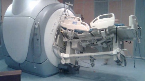
I’ve covered the problem of performing MRI on patients with external fixators. This is typically a problem that arises in head-injured patients with extremity or pelvic fixators for concomitant fractures.

MRI is an indispensable tool for the evaluation of head, spine, and soft tissue trauma. However, a great deal of effort is required to ensure that any patient scheduled for this test is “MRI compatible.” The fear is that any retained metallic fragments may move or heat up once the magnets are activated.

But what about trauma patients with external fixators? That is one big hunk of metal inserted deep into your patient. There are three major concerns:

  • Is the material ferromagnetic? If so, it will move when the magnets are activated and may cause internal injury. These days, many fixator sets are not ferromagnetic, avoiding this problem.
  • Can currents be induced in the material, causing heating? This is not much of a problem for small, isolated objects. However, external fixators are configured so that current loops can be created. The fluctuating magnetic fields can induce currents that, in turn, will heat the surrounding tissue. And thinner materials (narrow pins) result in more current and heating.
  • Will the metal degrade image quality?

Thankfully, there is a continuing trickle of evidence that is accumulating to give us some guidance. One paper from 2017 described a retrospective case series from four trauma centers. The authors performed MRIs on 38 patients with 44 external fixators. The adverse events they monitored for were catastrophic hardware pullout, thermal injury to the skin, field distortions that impaired the images, and damage to the magnet casing.

Twelve patients with 13 external fixators had MIR performed with the hardware inside the MRI bore, and 27 patients had the study with the fixator outside the bore. Most MRIs were performed to evaluate the cervical spine. There were no adverse events.

A recent Massachusetts General Hospital study involved a larger group (97 patients with 110 fixators). The fixators were located on the ankles, knee, femur, and pelvis. Most were performed on a 1.5T MRI, although a few were done on a 3T machine. Again, most scans were performed for head or cervical spine evaluation. Two of the 97 studies were terminated early due to patient discomfort. In both cases, the frame was outside the MRI bore.

The biggest challenge in our clinical practice is that there is no standard ex-fix configuration. Our orthopedic colleagues get to unleash their creativity, trying to devise the appropriate architecture to hold bones together so they can heal properly. This makes developing standardized guidelines regarding what can and can’t go into the scanner difficult.

We do know from clinical simulation studies that heating is influenced by ex-fix configuration. Increasing pin depth (thicker extremities) and closer pin spacing produces smaller temperature rises. For example, pins placed in a 15cm bar at a depth of 11cm produced a temperature rise of 2 degrees, but pins placed along a 30cm bar at a depth of 2cm showed a rise of 6 degrees.

However, a growing body of literature shows that the heating effects are relatively small and get smaller as the distance from the magnet increases. And non-ferromagnetic materials move very little, if at all, and do not interfere with the image. So as long as nonferromagnetic materials are used, the patients are probably safe as long as basic principles are adhered to:

  • Other diagnostic options should be considered and/or exhausted prior to using MRI.
  • Informed consent must be obtained, explaining that the potential risks are not completely understood.
  • The fixator must be tested with a handheld magnet so that all ferromagnetic components can be identified and removed.
  • All traction bows must be removed.
  • Ice bags or cooling packs should be placed at all skin-pin interfaces.
  • The external fixator should remain at least 7cm outside the bore at all times, if possible. If any portion must be inside the bore, monitoring efforts should be stepped up even more.

Bottom line: MRI of patients with external fixators can be safely accomplished. Consult your radiologists and physicists to develop a policy that is specific to the scanners used at your hospital. 

References:

  1. Magnetic Resonance Imaging of Trauma Patients Treated With Contemporary External Fixation Devices: A Multicenter Case Series. Journal of Orthopaedic Trauma, 31 (11), e375-e380. doi: 10.1097/BOT.0000000000000954.
  2. Magnetic Resonance Imaging of Trauma Patients Treated With Contemporary External Fixation Devices: A Multicenter Case Series. J Orthop Trauma. 2017 Nov;31(11):e375-e380. doi: 10.1097/BOT.0000000000000954. PMID: 28827510.


What Is: Lunchothorax?

Here’s an operative tip for trauma professionals who find themselves in the OR. Heard of “lunchothorax?” I’m sure most of you haven’t. The term originated in a 1993 paper on the history of thoracoscopic surgery. It really hasn’t been written about in the context of trauma surgery, though.

Lunchothorax is an empyema caused by pleural contamination in patients with concomitant diaphragm and hollow viscus injury. This most commonly occurs with penetrating injuries to the left upper quadrant and/or left lower back. The two penetrations tend to be in close proximity (diaphragm + stomach), but may occasionally be further away (diaphragm + colon).

One of the earlier papers describing the correlation of gastric injury and empyema was written by one of my mentors, John Weigelt. Although gastric repair is usually simple and heals well, his group did note a few severe complications. Of 243 patients with this injury, 15 developed ones that were considered severe, and 10 of those were empyema! What gives?

It turns out that the combination of gastric contents and pleural space is not a good one. It’s not really clear why this is. Is it bacterial? The acid? Undigested food? I’ve seen cases with what I would consider minimal contamination go on to develop a nasty empyema. This is also borne out in a National Trauma Databank review from 2009. It looked at complications in patients with a diaphragm injury and found that a gastric injury increased the probability of empyema by 3x. Interestingly, there was no increased risk of empyema with a concomitant colon injury.

Bottom line: Lunchothorax, or empyema after even minimal contamination from a hollow viscus, is a dreaded complication of thoraco-abdominal penetrating injury. Any time the stomach and diaphragm are violated, I recommend thoroughly irrigating the chest. It’s probably a good idea for concomitant colon injury as well, but there’s less literature support.

This can be done through the diaphragm injury if it is large enough, or through a chest tube inserted separately. Most of the time, you’ll be placing the chest tube anyway because the pleural space has been violated via the abdomen. In either case, copious lavage with saline is recommended to clear all particulate material, with a few extra liters just for good measure. There’s no data on use of antibiotics, but standard perioperative coverage for the abdominal injuries should be sufficient if the lavage was properly performed.

References:

  • The history of thoracoscopic surgery. Ann Thoracic Surg 56(3):610-614, 1993.
  • Penetrating injuries to the stomach. SGO 172(4):298-302, 1991.
  • Risk factors for empyema after diaphragmatic injury: results of a National Trauma Databank analysis. J Trauma 66(6):1672-1676, 2009. 

Closing Velocity And Injury Severity

Trauma professionals, both prehospital and in trauma centers, make a big deal about “closing velocity” when describing motor vehicle crashes.  How important is this?

So let me give you a little quiz to illustrate the concept:

Two cars, of the same make and model, are both traveling on a two lane highway at 60 mph in opposite directions. Car A crosses the midline and strikes Car B head-on. This is the same as:

  1. Car A striking a wall at 120 mph
  2. Car B striking a wall at 60 mph
  3. Car A striking a wall at 30 mph

2010-saab-9-5-head-on-crash-test_100313384_m1

The closing velocity is calculated by adding the head-on components of both vehicles. Since the cars struck each other exactly head-on, this would be 60+60 = 120 mph. If the impact is angled there is a little trigonometry involved, which I will avoid in this example. And if there is a large difference in mass between the vehicles, there are some other calculation nuances as well.

So a closing velocity of 120 mph means that the injuries are worse than what you would expect from a car traveling at 60 mph, right?

Wrong!

In this example, since the masses are the same, each vehicle would come to a stop on impact because the masses are equal. This is equivalent to each vehicle striking a solid wall and decelerating from 60 mph to zero immediately. Hence, answer #2 is correct. If you remember your physics, momentum must be conserved, so both of these cars can’t have struck each other at the equivalent of 120 mph. The injuries sustained by any passengers will be those expected in a 60 mph crash.

If you change the scenario a little so that a car and a freight train are traveling toward each other at 60 mph each, the closing velocity is still 120 mph. However, due the the fact that the car’s mass is negligible compared to the train, it will strike the train, decelerate to 0, then accelerate to -60 mph in mere moments. The train will not slow down a bit. For occupants of the car, this would be equivalent to striking an immovable wall at 120 mph. The injuries will probably be immediately fatal for all.

Bottom line: Closing velocity has little relationship to the injuries sustained for most passenger vehicle crashes. The sum of the decelerations of the two vehicles will always equal the closing velocity. Those injuries will be consistent with the change in speed of the vehicle the occupants were riding, and not the sum of the velocities of the vehicles. 

Artificial Platelets Under Development!

Uncontrolled bleeding is the bane of trauma professionals everywhere. Early in a resuscitation, we focus on identifying potential sources. We’ve developed numerous techniques for plugging them up. And we have processes in place to replace the blood that’s been lost.

Unfortunately, blood products are a perishable item. Packed red blood cells have a typical shelf-life of 42 days. Whole blood lasts only 21-35 days. Plasma is only suitable for up to five days once thawed. However, it can be frozen and used only when needed.

Platelets are another short-lifespan product, typically lasting only five days. This is a major reason for the relative lack of availability, especially at smaller hospitals. Unfortunately, freezing them or attempting cold storage renders them less active. For this reason, the platelet shortage persists.

As you know, platelets are fragments of cells produced by the bone marrow that have a major function in hemostasis. They bind to injured surfaces of disrupted blood vessels. Seconds later, they become activated and begin to clump with other platelets. They also release factors that result in fibrin deposition, creating a clot that helps stop bleeding.

Researchers have been trying to develop artificial blood substitutes for decades. I remember reading about rat experiments using these products in the 1980s. Unfortunately, they remain experimental to this day.

I found a recent article describing recent work on artificial platelets that piqued my interest. It was published by the biomedical engineering groups at North Carolina State University and UNC Chapel Hill. They used nanoparticles made of an ultrasoft microgel that were similar in size and shape to natural platelets. Fibrin-binding antibody fragments were embedded on the surface. These were selected to target only activated fibrin and not circulating fibrinogen.

Source: Science Translational Medicine

The groups devised a rat and pig trauma model by creating a liver laceration and then infusing varying doses of the artificial platelets (AP). Postmortem analysis of the wounds showed:

  • The APs did home in on the injured sites and were found in the injured areas
  • There was increased fibrin deposition at the wound site when compared to saline controls
  • Less bleeding was seen in the animals that received the APs vs saline
  • No significant deposition of APs was found in other tissues
  • The APs were excreted in the urine of the animals

Bottom line: This is very exciting, if preliminary, work. These artificial platelets are relatively easy to produce and can be frozen or stored at room temperature for extended periods. They appear harmless to the animals and decrease bleeding from the liver injury.

I am still somewhat cautious in my assessment. This same excitement was present 40 years ago in the early years of artificial hemoglobin solutions. And look where we are now. But, fingers crossed, there may be a solution to our chronic platelet shortage at some point in the future.

Reference: Ultrasoft platelet-like particles stop bleeding in rodent and porcine models of trauma. Sci Transl Med. 2024 Apr 10;16(742):eadi4490. doi: 10.1126/scitranslmed.adi4490. Epub 2024 Apr 10. PMID: 38598613.