EAST 2017 #9: Geographic Location and Fatal Car Crashes

Trauma resources (centers and helicopter services) are not geographically evenly distributed across the US. The East and West coasts are saturated with resources, maybe overly so. At the other extreme, some northern states (Alaska and the north central US) have very few trauma resources, and injured patients may have to travel several hundreds of miles to get definitive trauma care.

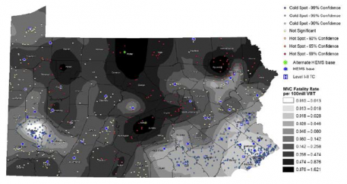
The group at the University of Pittsburgh looked at trauma resource distribution across the state of Pennsylvania, and matched that with geographical data from fatal car crashes over a two  year period. They used some special statistical tools to analyze this type of data, and reported their findings in a format that will be unfamiliar to many: fatalities per 100 million vehicle miles traveled (VMT).

Here are the factoids:

  • 863 fatal crashes occurred during the study period, killing 884 people
  • The median fatality rate for the state was .187 per 100 million VMT
  • Fatality hotspots became very apparent in areas farther from trauma system resources (TSR) (see map below, dark areas are bad)
  • The fatality rate increased significantly by 0.01 per 100 million VMT for each mile farther away from any TSR.
  • If just 2 helicopters had been relocated from trauma centers to high fatality regions, the overall fatality rate could have been reduced by 12%, in theory

Bottom line: This novel way of looking at injury data confirms what we all knew or suspected: injuries occurring farther away from trauma resources may lead to higher mortality and disability. And knowledge is power. If we can see it, we can do something about it. This type of analysis should be done on regional, state, and national levels to help us better serve our patients.

Questions and comments for the authors/presenters:

  1. Be able to describe your statistics simply
  2. How did you deal with data from the border areas of the state? Did you include trauma resources from adjacent states in your analysis?
  3. You mention “county-level” factors in adjusting mortality rates for distance. What were these?
  4. This is a novel way of approaching system planning. Nice job!

Click here to go the the EAST 2017 page to see comments on other abstracts.

Related post:

Reference: Distance matters: effect of geographic trauma system resource organization on fatal motor vehicle collisions. Paper #3, EAST 2017.

EAST 2017 #8: When Is “Mild TBI” Not So Mild?

Traumatic brain injury (TBI) is very common, with the majority falling into the “mild” category. This is usually defined as patients with injury to the head and a GCS of 13-15. These uncomplicated patients are frequently discharged from the emergency department, or undergo only a brief evaluation if admitted for other reasons.

The group at Shock Trauma focused on a less appreciated subset of mild TBI patients, those whose condition is a little more complicated. Specifically, these are patients with GCS 13-14 with positive findings on head CT leading to a calculated abbreviated injury score (head) of > 2, and some persistence of their symptoms while in the hospital. At many hospitals (including my own), these patients receive an inpatient TBI evaluation. But if they pass this initial screening, they are not consistently referred for any outpatient TBI followup.

Are these mild, complicated TBI patients (mcTBI) unique? Do they behave the same as the uncomplicated ones? The research group performed a prospective study on patients who sustained an mcTBI over a 4 month period.  They excluded patients with mental illness, dementia, and non-English speaking and homeless patients. They tried to contact patients up to three times after discharge to administer several standard tests and determine if they had any specific residual symptoms.

Here are the factoids:

  • Of the 142 patients with mcTBI during the study period, there was substantial attrition over time, with only 25 remaining at 6 months and 10 at one year
  • 64% of patients who responded at 6 months remained symptomatic. Depression, dizziness, and a feeling of impaired health were common.
  • 80% of patients still described symptoms at one year. The same complaints were most common, and some required changes in activities of daily living or assistive devices.

Bottom line: Although small and fraught with the usual problems in long-term tracking of urban trauma patients, this study is eye-opening. We too often dismiss “mild TBI” and being almost nothing, even in patients with positive findings on head CT. This work suggests that we are underestimating the needs of those patients. The authors used this data to design longer-term care processes for this subset of patients. Other centers should follow suit to make sure these patients’ post-injury needs are better met.

Questions and comments for the authors/presenters:

  • Describe the possible biases that patient selection and attrition may have had on the study
  • What type of TBI screening do you use in the hospital?
  • Given that a number of assessments were administered over the phone, I look forward to hearing some of the other details not listed in the abstract
  • Was there any correlation between specific CT findings and later symptoms?
  • Provide details of your long-term care programs for these patients
  • I enjoyed this thought provoking abstract!

Click here to go the the EAST 2017 page to see comments on other abstracts.

Related posts:

Reference: Mild TBI is not ‘mild’… survivors tell their complicated stories. Quick Shot #3, EAST 2017.

EAST 2017 #7: Pigtail vs Chest Tube – Does Size Matter?

I’ve been somewhat old school when it comes to chest tubes. Unlike some, I don’t believe that you have any control of where a chest tube goes if you are placing it in a closed chest. Only in the OR with an open one. And I’ve got plenty of x-rays to prove it.

And I used to think that chest tube size mattered when dealing with hemothorax. In theory, you need a big tube to get clots out, right?

Well, maybe not! The trauma group at the University of Arizona Tucson has previously done work on using 14 French pigtail catheters in lieu of a full-size tube. They will be presenting their extended experience with this concept at EAST 2017.

They have maintained a prospectively collected database of information on trauma patients with chest tubes for many years. This study focused only on those who had blood in their chest, either hemothorax (HTX) or hemopneumothorax (HPTX). They also looked at trends in their selection of chest drain tubes.

Here are the factoids:

  • Nearly 500 patients were treated with a tube for HTX or HPTX during the 7 year study period, 2/3 with a chest tube and 1/3 with a pigtail
  • Pigtails had more fluid drain initially (430cc vs 300cc, significant), and 1 less treatment day (4 vs 5, also significant)
  • Failure rate and insertion-related complications were the same (about 22% and 6%, respectively)
  • The group found that their use of pigtails steadily and significantly increased over the years

Bottom line: I’m coming around. The literature does appear to be tilting toward smaller tubes, and this longer-term study helps confirm that. How can this be? Although this is speculation on my part, it probably has to do with the fact that any size tube will drain liquid blood. And probably no size of tube will successfully get all the clot out. 

And certainly, smaller tubes are much better tolerated and do not require the degree of sedation that a mega-tube does. The authors suggest that a multi-center trial should be carried out to confirm this. For my part, I’m going to review the literature we have to date and consider modifying my own chest tube policy (see links below).

Questions and comments for the authors/presenters:

  • Where did you typically insert the pigtails? Anterior chest or classic chest tube position? Was it consistent?
  • Was/is the selection of tube type an attending surgeon specific choice, or did you implement a policy to direct them?
  • Did patient injury pattern or body habitus have any part in tube selection?
  • What about removal failures? That is, how many had to have a tube replaced, and how many went on to require VATS or other surgical procedure for drainage?
  • I enjoyed this provocative paper!

Click here to go the the EAST 2017 page to see comments on other abstracts.

Related posts:

Reference: A prospective study of 7-year experience of using percutaneous 14-French pigtail catheter for traumatic hemothorax at a Level I trauma – size still does not matter. Quick Shot #4, EAST 2017.

EAST 2017 #6: FAST Exam After Rolling to the Right

The FAST exam is an integral part of trauma evaluation. Even after experience and credentialing of providers, there tends to be some variability in performance. This is especially true when the abnormal findings (or amount of fluid present) is relatively small.

Can we improve this by doing something as simple as using gravity to help? When the patient is supine, fluid tends to pool in the pelvis, where interpretation is a little more complicated.  The surgery program at Guthrie/Packer Hospital created a small pilot study to see if they might improve the sensitivity of FAST by rolling patients to their right briefly, before returning to the supine position and performing the exam.

They enrolled seven participants who were already undergoing peritoneal dialysis (PD), so there was easy access to the peritoneal cavity for administration of known amounts of free fluid. First, each patient was drained of any residual dialysate via their PD catheter. They then underwent a baseline FAST exam. Next, they were placed in the right lateral decubitus position for 30 seconds, then placed supine again and the FAST was repeated. Each patient then had 50cc of dialysate infused, and the process was repeated until a positive FAST was obtained.

Here are the factoids:

  • Of the seven patients recruited, one was excluded because the initial FAST was equivocal due to body habitus and polycystic kidney disease
  • A maximum of 3 aliquots were given (150cc max)
  • Two patients became positive after right side down before any additional fluid was infused
  • None of the four remaining patients had a positive FAST after infusion of any aliquot in the supine position
  • All four became positive after the right side down maneuver,  two after 50cc, one after 100cc, and one after 150cc

Bottom line: The authors conclude that this may be a valuable technique to help detect smaller quantities of fluid than we normally do. I’m not so sure. First, it’s a tiny study in a patient group that is very different from trauma. And it’s impossible to quantify how much dialysate was left after initial drainage of the PD catheter. Finally, we know that FAST can’t “see” small quantities of fluid, but we have constructed our management algorithms around this fact. So we have a good idea of when we should do further imaging or run off to the operating room. Making this test more sensitive may skew these practice guidelines toward doing more (and potentially unneeded) imaging and surgery.

Questions and comments for the authors/presenters:

  • Did you record the volumes and administration times of dialysate given prior to the study? This may correlate with the initial positives and volumes needed to give a positive result.
  • Similarly, did you look at BMI and body habitus to see if there might be a correlation?
  • Are you planning any type of followup study, as you suggested in the abstract?

Click here to go the the EAST 2017 page to see comments on other abstracts.

Related posts:

Reference: Can we be faster? FAST examination after rolling to the right dramatically increases sensitivity. Quick Shot #7, EAST 2017.

EAST 2017 #5: Subarachnoid Hemorrhage, Neurosurgical Consults, and Repeat Head CT

Neurosurgical involvement in the management of simple traumatic brain injury (TBI) has been slowly dwindling over the past several years. This is the result of the general consensus that very few of these patients progress to need neurosurgical procedures.

A group at Wright State University in Dayton sought to define the progression of one specific finding in TBI, the subarachnoid hemorrhage (SAH). Secondarily, the wanted to determine if a neurosurgery consultation was warranted in these patients.

They performed a five year retrospective review of their registry data, identifying patients with both mild TBI (GCS 13-15) and SAH. They excluded patients with any other brain lesion on CT.

Here are the factoids:

  • 301 patients were enrolled during the 5 year period
  • All had a neurosurgical consultation
  • Time between the initial CT and a followup scan was about 11 hours
  • 91% showed stable or resolving SAH on the followup scan
  • 9% showed a worsening SAH or additional lesions on the repeat scan

Bottom line: The authors conclude that initial neurosurgical consultation is not needed, since only 9% of patients have worrisome findings on repeat CT. They do, however, recommend that the practice of repeat scanning be continued because of this same number.

Our trauma service looked at this issue a year ago, and determined that most of these lesions either do not progress, or never require any intervention if they do, with a few notable exceptions. For that reason, we abandoned both neurosurgical consultation and repeat CT scans for patients with non-aneurysmal SAH, a single parenchymal hemorrhage, or linear skull fractures. We continue to do both for patients with epidural and/or subdural hemorrhage. You can download a copy of this protocol here.

Questions and comments for the authors/presenters:

  • Did you look at platelet count or INR in the study. Were patients excluded based on abnormal values?
  • Did every patient get a repeat scan?
  • Break down the lesions in the 9% of patients who had some sort of progression or new finding. Did you see any common themes (age, chronic alcohol use, etc.)?
  • Did you encounter any patients with “non-central SAH”, that might indicate an aneurysm? How were they dealt with?
  • How has or will your trauma service change its practice based on your findings.

Click here to go the the EAST 2017 page to see comments on other abstracts.

Related posts:

Reference:   Management of subarachnoid hemorrhage (SAH) by the trauma service: are repeat CT scanning & routine neurosurgical consultation necessary? Poster #16, EAST 2017.