When To Remove a Chest Tube

Chest tubes are needed occasionally to help manage chest injuries. How do you decide when they are ready for removal?

Unfortunately, the literature is not very helpful in answering this question. To come up with a uniform way of pulling them, our group looked at any existing literature and then filled in the blanks, negotiating criteria that we could all live with. We came up with the following.

Removal criteria:

  • No (or a minimal, stable) residual pneumothorax
  • No air leak
  • Less than 150cc drainage over the past 3 shifts. We do not use daily numbers, as it may delay the removal sequence. We have moved away from the “only pull tubes on the day shift” mentality. Once the criteria are met, we begin the removal sequence, even in the evening or at night.

Removal sequence:

  • Has the patient ever had an air leak? If so, they are placed on water seal for 6 hours and a followup AP or PA view chest x-ray is obtained. If no pneumothorax is seen, proceed to the next step.
  • Pull the tube. Click here to see a video demonstrating the proper technique.
  • Obtain a followup AP or PA view chest x-ray in 6 hours.
  • If no recurrent pneumothorax, send the patient home! (if appropriate)

Click here to download the full printed protocol.

Off-Label Use of the Foley Catheter

Foley catheters are a mainstay of medical care in patients who need control or measurement of urine output. Leave it to trauma surgeons to find warped, new ways to use them!

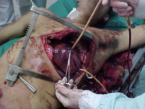
Use of these catheters to tamponade penetrating cardiac injuries has been recognized for decades (see picture, 2 holes!). Less well appreciated is their use to stop bleeding from other penetrating wounds.

foleyinheart

Foley catheters can be inserted into just about any small penetrating wound with bleeding that does not respond to direct pressure. (Remember, direct pressure is applied by one or two fingers only, with no flat dressings underneath to diffuse the pressure). Arterial bleeding, venous bleeding or both can be controlled with this technique.

In general, the largest catheter with the largest possible balloon should be selected. It is then inserted directly into the wound until the entire balloon is inside the body. Inflate the balloon using saline until firm resistance is encounted, and the bleeding hopefully stops. Important: be sure to clamp the end of the catheter so the bleeding doesn’t find the easy way out!

Use of catheter tamponade buys some time, but these patients need to be in the OR. In general, once other life threatening issues are dealt with in the resuscitation room, the patient should be moved directly to the operating room. In rare cases, an angiogram may be needed to help determine the type of repair. However, in the vast majority of cases, the surgeon will know exactly where the injury is and further study is not needed. The catheter is then prepped along with most of the patient so that the operative repair can be completed.

Related posts:

Syncope Workup in Trauma Patients

Syncope accounts for 1-2% of all ED visits, and is a factor in some patients with blunt trauma, especially the elderly. If syncope is suspected, a “syncope workup” is frequently ordered. Just what this consists of is poorly defined. Even less understood is how useful the syncope workup really is.

Researchers at Yale retrospectively looked at their experience doing syncope workups in trauma patients. They were interested in seeing what was typically ordered, if it was clinically useful, and if it impacted length of stay. 

A total of 14% of trauma patients had syncope as a possible contributor to their injury. The investigators found that the following tests were typically ordered in these patients:

  • Carotid ultrasound (96%)
  • 2D Echo (96%)
  • Cardiac enzymes (81%)
  • Cardiology consult (23%)
  • Neurology consult (11%)
  • EEG (7%)
  • MRI (6%)

Most of this testing was normal. About 3% of cardiac enzymes were abnormal, as were 5% of carotid imaging and 4% of echocardiograms. 

Important! Of the patients who underwent an intervention after workup, 69% could have been identified based on history, physical exam, or EKG and did not depend on any of the other diagnostic tests.

Conclusion: Syncope workup is not needed routinely in trauma patients with syncope as a contributing factor. Need for intervention can usually be determined by history, exam and EKG performed in the ED. In this study, $216,000 in excess costs would have been saved!

Reference: Routine / protocol evaluation of trauma patients with suspected syncope is unnecessary. Davis, et al, Yale University. Presented at the 23rd Annual Scientific Assembly of the Eastern Association for the Surgery of Trauma, January 2010.

EMS: Which Field Airways Work The Best?

Oral endotracheal intubation is the gold standard when a field airway is needed. However, they are not always possible due to protocol, training, patient anatomy or specific injuries. To allow airway support in these situations, a number of techniques and devices have been developed. The problem is, do we really know which one(s) are best?

To try to answer this question, a huge meta-analysis of all the English literature with information on success rates for these techniques was carried out. Over 2000 papers were identified, and they were narrowed down to 35 studies involving over 10,000 patients. 

The success rates that they identified were as follows:

  • King LT airway – 96.5%
  • Esophageal Obturator / Esophageal Gastric Tube Airway – 92.6%
  • Surgical cricothyroidotomy – 90.5%
  • Laryngeal mask airway (LMA) 87.4%
  • Combitube – 85.4%
  • Pharyngeotrachael laryngeal airway (PTLA) – 82.1%
  • Needle cricothyroidotomy – 65.8%

The Bottom Line: The King airway has the highest success rate of the alternative airway devices, although there was less data available and the effectiveness of ventilation has not been worked out yet. The best percutaneous rescue airway was the surgical crich.

Reference: A Meta-Analysis of Prehospital Airway Control Techniques Part II: Alternative Airway Devices and Cricothyrotomy Success Rates. Prehospital Emergency Care 14(4):515-530, Oct-Dec 2010.

Trauma Factoid: What Cars Were Doing When Involved In A Fatal Crash?

What were cars doing when they got involved in a fatal crash?

  • 69% – going straight ahead
  • 15% – negotiating a curve
  • 6% – turning left
  • 4% – not known
  • 2% – passing
  • 2% – merging or changing lanes
  • 1.5% – stopping, slowing or starting in a traffic lane
  • 0.8% – turning right

Bottom line – most fatal crashes occur when moving straight ahead, but look out when turning left!!

Reference: NHTSA