Tag Archives: chest tube

Best Of EAST 2024 #4: Chest Tube Irrigation Prevents Retained Hemothorax

One of the potential complications of chest trauma causing hemothorax is the retained hemothorax. In most patients, retained blood slowly lyses and is reabsorbed. But a few do not, and scarring can occur that entraps the lung and interferes with pulmonary function. This can ultimately require a VATS or thoracotomy to resolve.

Several protocols have been developed to try to prevent a retained hemothorax. They include the use of lytics or an early VATS procedure. The group at the Medical College of Wisconsin performed a trial of thoracic cavity irrigation and compared the outcomes with patients who did not undergo irrigation. This was a single-center retrospective study performed over five years.

This appears to have been a common practice at this institution. Patients undergoing chest tube placement for hemothorax received irrigation of the chest cavity immediately after placement. The study excluded patients with chest tubes placed in other hospitals, tubes placed late (after 24 hours), or patients who had a chest procedure within 6 hours.

Here are the factoids:

  • A total of 370 patients were enrolled, and 225 (61%) received irrigation
  • Demographics of the groups were the same, with the exception that the irrigation group contained more patients with penetrating injury and fewer patients with flail chest
  • Use of irrigation was associated with significantly less incidence of retained hemothorax (10% vs 21%) or need for VATS (6% vs. 19%)
  • Chest tube duration (4 vs 6 days) and hospital length of stay 8 vs 10 days) were also significantly shorter

The authors concluded that irrigation prevents retained hemothorax and decreases the need for surgical intervention.

Bottom line: Well, this was a new one for me. The only prior study I could find was published in 2022 by a group at the University of Nevada at Las Vegas. They irrigated 82 of 198 patients undergoing chest tube placement. They noted a decrease in hospital, ICU, and ventilator days.

This study looks at something far more practical: interruption of the development of a complication. Although still a relatively small and single-institution study, it was well done and could easily detect statistical significance. 

The presenter should be prepared to discuss what impact the mechanism of injury (penetrating, flail chest) may have had on their results and the exact technique they used. How much fluid, what type, and how it was drained are all important questions to discuss.

This is a fascinating abstract indeed. If the presentation answers the questions, centers should consider updating their chest tube management algorithms.

References:

  1. Thoracic cavity irrigation prevents retained hemothorax and decreases surgical intervention in trauma patients. EAST 2024, Podium paper #17.
  2. The Volume of Thoracic Irrigation Is Associated With Length of Stay in Patients With Traumatic Hemothorax. J Surg Res. 2022 Nov;279:62-71.

Best Of AAST #3: When To Place A Chest Tube For Hemothorax

There is an art to deciding when to place a  chest tube for either hemothorax or pneumothorax. For the most part, the trauma professional examines the imaging and then uses some unknown internal metric to declare that it is “too big.” Then it’s time to insert some type of chest drain.

There have been attempts over the years to make this decision more quantitative. One of the better-known ones is the 2-cm rule for pneumothorax. If the distance from the chest wall to the lung on the chest x-ray is >2cm, it is “too big.”

But what about hemothorax? The Medical College of Wisconsin trauma group performed a retrospective review of 391 patient charts to test a new 300cc rule defining when a hemothorax is “too big.” This guideline was implemented in 2018-2019, and patients presenting before implementation were compared to those arriving after.

The 300cc threshold is determined by using Mergo’s formula for calculating the volume of a square prism. Obviously, this requires a CT scan for calculation, so patients who had a tube placed before scanning or did not have one were excluded. They were also excluded from the study if their pneumothorax met the 2-cm rule. The authors studied how many patients could be observed, how many needed tube drainage, observation failure, and later need for a VATS procedure or thoracotomy.

Here are the factoids:

  • About 60% of the study group was admitted after the new criteria were implemented, and both groups were demographically similar
  • After implementation, the number of patients that were just observed increased significantly from 52% to 71%
  • Of course, this means that the number of chest tubes inserted was significantly less (42% vs. 61%)
  • There was no difference in observation failure (delayed placement of a tube), 18% vs. 24%
  • There were also no differences in pulmonary complications, 30-day readmissions, or 30-day mortality
  • The average ICU and hospital length of stays were significantly shorter as well

The authors concluded that implementing their 300cc guidelines correlated with decreased length of stay and no increase in failure or complication rates.

Bottom line: Although this is a relatively small series, the differences between the groups quickly achieved significance. There are three major questions that I have. First, how was the 300cc threshold arrived at? Was this borne of clinical judgment, or did some previous work suggest it?

My next question has to deal with the accuracy of the volume calculation. Mergo’s formula was used to determine the volume of a rectangular solid. As we all know, hemothoraces and pneumothoraces are not cubes. They can be very irregular and influenced by patient position. However, I did find a paper from the University of Florida that found the correlation coefficient between the volume calculated by Mergo’s formula vs. using 3-D software estimation was 0.9, which is excellent. So this approximation appears to be a very good one.

Finally, using the 300cc rule is predicated on getting a CT scan. Does every patient need a chest CT? Part of the resuscitation process for major trauma involves obtaining a chest X-ray. The obviously large hemothorax can justify inserting a chest tube at that point. But the reality is that most of these patients do go on to chest CT, so this is a minor change in practice for most. 

Although I love to see confirmatory studies before practice changes, this one study can lead us to change our practice guidelines now. It is a relatively minor one and will allow us to avoid placement of a few more chest tubes and to shave off a few days of hospital stay. The logical follow-up study for the authors is to extend the post-discharge window for complications to 60 or 90 days to ensure that delayed procedures were not required in the observation group.

References:

  • Implementing the 300-cc rule safely decreases chest tube placement in traumatic hemothorax. AAST 2023 Plenary paper #22.
  • New formula for quantification of pleural effusions from computed tomography. J Thorac Imaging. 1999 Apr;14(2):122-5. 


Chest Tube Based On Pneumothorax Size

How big is too big? That has been the question for a long time as it applies to pneumothorax and chest tubes. For many, it is a math problem that takes into account the appearance on chest x-ray, the physiology of the patient, and their ability to tolerate the pneumothorax based on any pre-existing medical conditions.

I first wrote about this paper when it was just an abstract for last year’s AAST meeting. Apparently, it passed peer review muster. It has just been published in the Journal of Trauma. The numbers have changed a little bit, so I’ll update my analysis accordingly.

The group at Froedtert in Milwaukee has been trying to make the decision to place a chest tube a bit more objective. They introduced the concept of CT based size measurement using a 35mm threshold at the AAST meeting three years ago. Read my review here. My criticisms at the time centered around the need to get a CT scan for diagnosis and their subjective definition of a failure requiring chest tube insertion. The abstract never did make it to publication.

The authors are back now with a follow-on study. This time, they made a rule that any pneumothorax less than 35mm from the chest wall would be observed without tube placement. The performed a retrospective review of their experience and divided it into two time periods: 2015-2016, before the new rule, and 2018-2019, after the new rule. They excluded any chest tubes inserted before the scan was performed, those that included a sizable hemothorax, and patients placed on a ventilator or who died.

Here are the factoids:

  • There were 99 patients in the early period and 167 in the later period
  • Chest tube use significantly declined from 28% to 18% between the two periods. These numbers are 8% higher than were described in last year’s abstract.
  • Observation rates without a chest tube increased from 85% to 95% after implementation of the new guideline
  • There was no difference in length of stay, inpatient failure rate, complications, or death
  • The most common inpatient failure was due to development of a new hemothorax. However, there was an almost identical number of failures of “unclear” etiology. This is troublesome but part and parcel for such a retrospective study.
  • Two patients were readmitted within 30 days for a pulmonary complication (one empyema, one readmission at 3 days after discharge for dyspnea due to pneumothorax)
  • Patients in the later group were 2x more likely to be observed (by regression analysis)

The authors concluded that the 35mm rule decreased unnecessary chest tube insertion while maintaining patient safety.


Bottom line: I still have a few issues with this paper and the authors’ preceding series of abstracts. First, decision to insert a chest tube required a CT scan in a patient with a pneumothorax. This seems like extra radiation for patients who may not otherwise fit any of the usual blunt imaging criteria. And, like their 2018 and 2021 abstracts, there are no objective criteria for failure requiring tube insertion. So it is difficult to gauge compliance when insertion for failure is somewhat based on the whims of the individual surgeon.

What this abstract really shows is that compliance with the new rule increased, and there were no obvious complications from its use. The other numbers (chest tube insertions, observation failure) are just too subjective to learn much from. The most troubling issue is that the reason for 40% of failures was “unclear.” This is most likely due to the fact that the authors did not have objective guidelines for failure due to the retrospective nature of the study.

The numbers in this paper changed a little from last year’s abstract. The overall conclusions and meaning did not. It appears that 35mm is a reasonable threshold for pneumothorax size when contemplating inserting a chest tube. Unfortunately, this study relied entirely on CT scan. We don’t know if using a similar guideline for regular old chest x-ray is valid or not. 

What we still need is a good, prospective trial using an arbitrary guideline like 35mm pneumothorax as seen on chest x-ray or CT scan. And then, a clear definition of what defines a failure that requires tube insertion would be helpful. And at some point, we also need to know if a small tube or pigtail catheter is adequate for pure pneumothorax. Don’t get me started on that one!

Reference: The 35-mm rule to guide pneumothorax management: Increases appropriate observation and decreases unnecessary chest tubes. J Trauma 92(6):951-957, 2022.

Best Of EAST #5: Ultrasound vs Chest X-Ray After Chest Tube Removal

The chest is one of the most commonly injured body regions. Patients are frequently found to have either air or blood in the chest, and many require a chest tube (tube thoracostomy) for these conditions. There is an art to chest tube removal, and even in 2021, the best practice has not been fully worked out.

Some believe that pulling the tube during a breath hold is best. Others do this during full expiration. Most centers confirm an uneventful tube removal with a plain chest x-ray. But the time interval after removal varies considerably.

The trauma group at the University of Tennessee – Chattanooga examined the use of chest ultrasound as the confirmatory test for residual pneumothorax after removing a chest tube. They developed an institutional practice guideline requiring a trans-thoracic ultrasound performed by a first-year resident two hours after tube removal. The interns all completed a 30-minute standard ultrasound course for training prior to beginning the study.

Two hours after tube removal, an intern performed the ultrasound (US) and interpreted it. A chest x-ray (CXR) was then ordered and the results compared.

Here are the factoids:

  • A retrospective review of 46 patients was performed, but the inclusion criteria were not listed in the abstract
  • Eleven of the 46 (24%) had a residual pneumothorax on CXR, and the US detected it in 12 (26%)
  • Three patients had PTX on CXR, but not US
  • Four patients had PTX on US, but not CXR
  • None of the PTX were clinically significant, and none required tube reinsertion
  • Cost of care savings was projected to be $4,000 if chest x-ray was not needed

The authors concluded that bedside ultrasound was an acceptable alternative to chest x-ray, with decreased radiation exposure and cost.

Bottom line: This is an intriguing abstract. It shows us that there might be an alternative to the standard chest x-ray confirmation after chest tube removal. It’s a very small study, so non-inferiority can’t truly be established yet. The studies are complementary since each study misses a few pneumothoraces that the other picks up.

At this point, I wouldn’t recommend switching entirely to ultrasound until we have a larger series. But I bet we will be able to in the future. Ultimately, this could reduce radiation exposure (tiny anyway for a chest x-ray) and save a small amount of money. But it will reduce x-ray department resource usage, which may be very helpful for the hospital.

Here are my questions for the authors and presenter:

  • How did you select your patients? What were the selection criteria? How long did it take to accrue 46 patients? It’s important that all patients with a chest tube had the criteria applied, otherwise there is an opportunity for bias. We want to make sure that you didn’t inadvertently enroll only the patients for whom ultrasound works well.
  • How much of a burden was placed on the interns who did the exam? Was the ultrasound unit nearby? Or did they have to spend 30 valuable minutes rolling it to the floor and doing the study? Radiology department resource use needs to be balanced with intern resource utilization.
  • Why did you have such a high rate of residual pneumothorax after the tubes were pulled (about 25%). This seems a bit higher than what the literature reports.
  • What does your protocol require when a residual pneumothorax is found? Do you have to perform another study after an additional time interval to prove that it is not getting larger? Serial ultrasound exams? Another chest x-ray? Please show us your entire guideline.

I really enjoyed this paper. I’m looking forward to hearing the nitty gritty details during the presentation.

Reference: ULTRASOUND SAFELY REPLACES CHEST RADIOGRAPH AFTER TUBE THORACOSTOMY REMOVAL IN TRAUMA PATIENTS. EAST 25th ASA, oral abstract #9.

Practice Guideline: Chest Tube Management (Part 2)

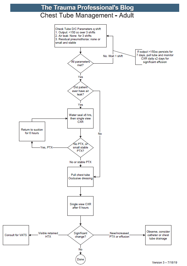
In my last post, I went over the rationale for developing a practice guideline for something as simple and lowly as chest tube management. Today, I’m posting the details of the guideline that’t been in use at my hospital for the past 15 years. I’ve updated it to reflect two lessons learned from actually using it.

Here’s an image of the practice guideline. Click to open a full-size copy in a new window:

Here are some key points:

  • Note the decision tree format. This eliminates uncertainty so that the clinician can stick to the script. There are no hedge words like “consider” used. Just real verbs.
  • We found that hospital length of stay improved when we changed the three parameters from daily monitoring to three consecutive shifts. We are prepared to pull the tube on any shift, not just during the day time. And it also allows this part of the guideline to be nursing driven. They remind the surgeons that criteria are met so we can immediately remove the tube.
  • Water seal is only used if there was an air leak at some point. This allows us to detect a slow ongoing leak that may not be present during our brief inspection of the system on rounds.
  • The American College of Surgeons Committee on Trauma expects trauma centers to monitor compliance with at least some of their guidelines. This one makes it easy for a PI nurse or other personnel to do so.
  • The first of the “new” parts of this guideline is: putting a 7 day cap on failure due to tube output greater than 150cc per three shifts. At that point, the infectious risks of keeping a tube in begin to outweigh its efficacy. Typically, a small effusion may appear the day following removal, then resolves shortly.
  • The second “new” part is moving to VATS early if it is clear that there is visible hemothorax that is not being drained by the system. Some centers may want to try irrigation or lytics, but the data for this is not great. I’ll republish my posts on this over the next two days.

Click here to download a copy of this practice guideline for adults.

Click here to download the pediatric chest tube practice guideline.