Galleries

Best Of: What Percent Pneumothorax Is It?

Frequently, radiologists and trauma professionals are coerced into describing the size of a pneumothorax seen on chest xray in percentage terms. They may something like “the patient has a 30% pneumothorax.”

The truth is that one cannot estimate a 3D volume based on a 2D study like a conventional chest xray. Everyone has seen the patient who has no or a minimal pneumothorax on a supine chest xray, only to discover one of significant size with CT scan.

Very few centers have the software that can determine the percentage of chest volume taken up with air. There are only two percentages that can be determined by viewing a regular chest xray: 0% and 100%. Obviously, 0% means no visible pneumothorax, and 100% means complete collapse. Even 100% doesn’t really look like 100% because the completely collapsed lung takes up some space. See the xray at the top for a 100% pneumothorax.

If you line up 10 trauma professionals and show them a chest xray with a pneumothorax, you will get 10 different estimates of their size. And there aren’t any guidelines as to what size demands chest tube insertion and what size can be watched.

The solution is to be as quantitative as possible. Describe the pneumothorax in terms of the maximum distance the edge of the lung is from the inside of the chest wall, and which intercostal space the pneumothorax extends to. So instead of saying “the patient has a 25% pneumo,” say “the pneumothorax is 1 cm wide and extends from the apex to the fifth intercostal space on an upright film.”

Technology: Real Time Cerebral Blood Flow Monitoring For TBI

Here’s a new toy that has recently received some funding from the US military. It allows real-time monitoring of cerebral blood flow. It may help identify flow problems from elevated intracranial pressure (ICP) or vasospasm early on, allowing prompt initiation of appropriate therapies to increase blood flow.

This device uses an array of ultrasound beams and locks onto the middle cerebral artery. It then continuously monitors blood flow and displays the result in real time. I predict that there will be a learning curve with this one, similar to near infrared monitoring of tissue perfusion. What’s a normal baseline? What kind of variation is considered “normal?” We’ll have to answer these questions before this tool is ready for prime time. Ultimately, it may allow noninvasive monitoring of ICP in the intensive care unit.

Credit: Physiosonics, Bellevue, WA.

Blood Transfusion With Component Therapy

About 40 years ago, blood banks started moving away from keeping whole blood and began separating it into components (packed cells, platelets, plasma, etc.) for more targeted use. For most uses, this is just fine. But what about trauma?

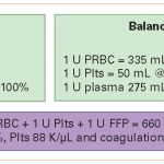
Trauma patients bleed whole blood. Doesn’t it make sense to give whole blood back? Much of our experience with massive transfusion is derived from our colleagues in the military. Two decades ago, the norm was to give 4 units of packed red cells or so, then give two units of plasma, and every once in a while slip in a bag of platelets. Our military experience seems to indicate that this 4:2:1 ratio is not optimal, and that something like 1:1:1 is better.

If you think about it, whole blood is already 1:1:1. Splitting it into components and then giving them back seems to be a lot of extra work (and expense) to accomplish the same thing as just giving a unit of whole blood. Plus it triples the exposure to infectious agents and antigens, since the components will usually come from three separate donors. Note that the data in the table above is true for fresh whole blood (not practical in civilian life); banked whole blood will lose some coagulation activity.

Is it time to think about supplying whole blood to trauma centers? And actually looking at whether the outcomes are better or not?

How To Manage TBI In Patients On Warfarin

We all know that the combination of traumatic brain injury (TBI) and warfarin can be dangerous. Here at Regions, we developed a reversal protocol a few years ago. However, we found that just having a list of preferred “antidotes” to give was not enough. The time factor is very important, and we found that we needed to ensure prompt use of these medications when indicated.

So we added features that ensured timely response and reversal. You can download the protocol by clicking the image above or the link at the bottom of this post.

First, we recognized that any patient with a known or suspected TBI who was taking warfarin was at risk. If the initial GCS was <14, then a full trauma team activation is called. This gives the patient priority lab processing and immediate access to the CT scan. In addition, 2 units of thawed plasma are administered while in the resuscitation room. If the head CT is negative, plasma is stopped.

For patients with a GCS of 14 or 15, a “Code RED” is called, ensuring that an ED physician sees the patient immediately. A point of care INR is drawn and the patient is sent for stat head CT. If the head CT is negative with INR>2.5, the patient is admitted for observation and a repeat head CT is obtained 12 hours later. We have seen patients develop delayed hemorrhage when they have high INR.

We apply a restrictive set of criteria to determine if a patient may go home from the ED, which causes us to admit most for observation. And if they do have a positive CT, we use the algoritm listed below for comprehensive management and reversal.

Bottom line: Patients with any head trauma and an elevated INR are a walking time bomb. They need prompt assessment and reversal of their anticoagulation if indicated. Feel free to share your protocols here as well by posting a comment.

Download the full protocol; click here.

Related post: