Galleries

What The Heck Is It? Final Answer!

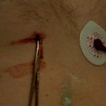
Yesterday, I posted the physical exam findings on this mystery object. A tiny puncture wound was present just to the right of the xiphoid on the lower chest wall, and a small sharp point was palpable.

Here’s how to deal with it:

Step 1. (Image 1) Don’t let it move or try to pull it out immediately! I didn’t want this thing to go in any further, or work it’s way out in case it was in the heart. I snapped a hemostat on the end of it for stabilization.

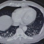
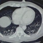
Step 2. (Images 2 and 3) Find out where it is exactly. You need to know what if any vital structures it may have pierced so you can plan for removal. In this stable patient, CT was the best option. If he had been unstable, it would have meant an immediate trip to the OR. Note how the object is within the chest wall, BUT it had penetrated at least as deep as the lung since a pneumothorax was present.

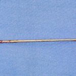
Step 3. (Image 4) Get it out! Off to the operating room for removal, just in case some unexpected bleeding or hemodynamic changes were to occur. After the patient was asleep, a chest tube was inserted. The object was a fine nail from a commercial nail gun mishap and pulled out easily with the hemostat. He was discharged two days later after the tube was pulled. 

Photo source: personal archive. Patient not treated at Regions Hospital

Concussion Testing: There’s An App For That!

Smart phone programmers are becoming more and more creative! The newest trauma app is geared toward helping the user identify individuals who have suffered a concussion. It can be used by parents, coaches or physicians to help identify a concussion at sporting events.

The app is a portable and convenient system for identifying concussions based on established sports medicine research. It queries the user for common signs of concussion, tallies the results of a simple balance test, and looks for other symptoms that suggestion the injury. The exam can also be administered serially to detect changes from baseline.

To get the most from this free app, the user must purchase an optional module for $4.99 that does a more in-depth physiologic and cognitive evaluation. A report can be emailed automatically to your physician, and he or she can then respond and send a message to your team to approve or deny continuing play.

The app is provided by SportSafety Labs LLC. The basic app is free, and the add-on is $4.99. It is published for the iPhone and iPad.

Bottom line: Expect more trauma-oriented apps geared toward a variety of problems in the near future!

To get more information on this app, click here.

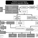
Algorithm For Clearing the Pediatric Cervical Spine

I previously wrote about a straightforward way to clear the cervical spine in children. Click here to see the article. Alfred I. DuPont Children’s Hospital has condensed their clearance technique into a relatively simple algorithm that can be used in conjunction with my previous tips.

Some notes on this algorithm:

  • Can be performed only by attending physicians or a trauma resident in consultation with the attending trauma surgeon
  • Clinical clearance alone may be carried out in select cases
  • If radiographs are required, cross-table lateral, anterior/posterior, and odontoid views should be obtained (age 8 and above, non-intubated)
  • Flexion / extension views should only be ordered in consultation with neurosurgery

Download a print version of the protocol here

Related post: How Do I Clear The Pediatric Cervical Spine?

Image and protocol courtesy of the Alfred I DuPont Children’s Hospital

Is It Really Safe To Observe Occult Pneumothorax?

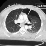
Occult pneumothorax is the most common incidental finding on CT imaging, occurring in 2% to 10% of trauma patients. By definition, an occult pneumothorax is a pneumothorax that is seen only on CT and not a conventional chest x-ray. When detected, the question that comes to mind is, will this patient need a chest tube?

The AAST conducted a trial encompassing the experience at 16 Level I and II trauma centers around the US. They looked at injury severity, specific chest injuries, ventilator settings if on positive pressure ventilation (PPV) and size of pneumothorax. The size was calculated by measuring the largest air collection along a line perpendicular to the chest wall (see image above). Failure of observation meant that a thoracostomy tube was placed.

The 2 year study looked at a total of 448 occult pneumothoraces that were initially observed. Key findings of the study were:

  • Injury severity was no different between failure and non-failure groups
  • There was a 6% failure rate overall
  • PPV alone was associated with an increased failure rate of 14%
  • Surgical intervention requiring PPV was not associated with an increased failure rate
  • Pneumothorax size > 7mm, positive pressure ventilation, progression of the pneumothorax, respiratory distress and presence of hemothorax were associated with failure.
  • Pneumothorax size was not entirely reliable for predicting failure, since patients with sizes as small as 5mm on PPV and 3mm not on PPV failed in this series

Bottom line: Most blunt trauma patients with an occult pneumothorax can be safely observed. A followup chest x-ray should be obtained to look for progression. If the patient progresses, is placed on PPV, has a hemothorax or develops respiratory distress, have a low threshold for inserting a drainage tube. Maximum pneumothorax size may predict failure when large, but it can still happen with very small air collections.

Related posts:

Reference: Blunt traumatic occult pneumothorax: is observation safe? – results of a prospective, AAST multicenter study. J Trauma 70(5):1019-1025, 2011.

CT image courtesy of Journal of Trauma