Tag Archives: TBI

Stroke And Fall VS Fall And Bleed

It’s like the old chicken and egg question. When dealing with head trauma and falls, which came first? Did the patient have a stroke and then fall down? Or did they fall and sustain some type of intracranial hemorrhage? And you may ask, does it make a difference? They are going to get a head scan anyway, right?

In my opinion, it makes a big difference! How often have you seen the following scenario? EMS is called to a house or nursing home for someone who has fallen. They notice some extremity weakness on one side and presume the patient is having a stroke. The emergency department is then notified that a stroke patient is inbound.

On arrival, the patient was rapidly assessed and whisked off to CT scan for a CT and angiogram, possibly with neurology present. My experience is that a majority of these scans is negative for CVA. And many are positive for some type of extra-axial hemorrhage like subdural or epidural blood from the real injury.

Unfortunately, something called anchoring bias is likely to occur in this situation. Everyone from the paramedics onward are moving along under the assumption that the patient has had a stroke. They stop considering the more common diagnosis of TBI and other potential injuries in the spine and torso. Even when the CT angiogram is found to be negative, it’s difficult for people to change gears. It then takes longer to address the subdural or epidural. The involved trauma professionals are less likely to activate the trauma team. And further evaluation of the chest, abdomen, and spine may be delayed or forgotten for a time.

Bottom line: In any case of a fall followed by neurological changes that could indicate stroke, always presume a serious TBI first! If EMS requests a stroke code, it should be changed to a trauma activation prior to patient arrival. This takes advantage of the odds (more in favor of TBI) and activates a team that is well versed in evaluating the entire patient. If no evidence of hemorrhagic stroke is present, the team will then order the brain CTA and involve the stroke team as necessary.

And for good measure, every one of these cases that does start as a stroke evaluation should be addressed by the trauma performance improvement process!

Best Of EAST #15: Prehospital TXA

The world is divided into trauma centers that are TXA believers and those that are TXA nonbelievers. It all depends on how one interprets the CRASH-2 data and subsequent studies. Then came CRASH-3 with TXA use for patients with TBI. This large study found improved survival in patients with mild to moderate head injury when given “early.”

The group at Oregon Health Science University tried to better define this concept of “early.” They examined early vs later administration of TXA in patients with moderate to severe TBI. Note that this degree of head injury is a bit different than CRASH-3 (mild to moderate in CRASH-3 and moderate to severe in this one). This was a multicenter trial that included patients with GCS < 12 and who were hypotensive with SBP < 90. Patients received either a 1g bolus followed by a 1g infusion over 8 hours, or a 2 g bolus only. The authors subdivided these patients into early administration (<45 minutes after injury) or late (45 minutes to 2 hours after injury).

Here are the factoids:

  • There were 354 patients in the early administration group and 259 in the late group
  • All outcomes, including 1 month and 6 month mortality and the extended Glasgow outcome scale were not significantly different between early and late groups (exact numbers were not given)
  • There was no difference in secondary complications between the groups (again, exact numbers or complication types were not given)

The authors concluded that there was no difference in outcomes in early vs later administration of TXA in these head injured patients. They suggest that patients can be given TXA anytime within two hours without loss of benefit.

Bottom line: Essentially, this ends up as a noninferiority study. The biggest question with this type of study is, do you have enough subjects to detect a significant difference? Taken to an extreme, let’s say you have 5 patients who receive a drug who are compared to 5 who did not for some mystery condition. Three who did not get the drug die (60% mortality), but only two who get it do (40% mortality). In relative terms, there was 33% decrease in mortality with the drug. But in absolute terms, it was one patient. Would anyone see this as a significant result with such small numbers?

But now multiply by a thousand, and 300 die without the drug and only 200 die who were given it. The relative difference is the same, but the absolute difference is beginning to look large and significant.

So the smaller study won’t meet the test of significance but the larger one will. The key question in the TXA study here is, do they have enough patients enrolled to show there is no real difference between the groups? I love doing back of the napkin power analyses, and I admit I certainly don’t have all the numbers and probabilities needed for a precise calculation. But the groups sizes in this study (354 vs 259) seem a bit small to achieve significance unless there are large disparities in outcomes. 

I certainly recognize that it’s just not possible to put all the relevant information for a research project into a four paragraph abstract. One would need to be able to submit 12 slide PowerPoint decks. So I’m sure more info will be available as I take in the presentation next Friday.

Here are my questions for the authors and presenter:

  • The study is nicely designed as a randomized, double-blind trial, but how did you blind one vs two doses? Did everyone get an infusion of something, TXA vs saline?
  • Why did you select 45 minutes as the cutoff for early vs late administration? Was this arbitrary or is it based some data?
  • Show us the power analysis that demonstrates the total number of patients in the study is sufficient to show us true non-significance in your results.
  • And I’m sure you will show the actual survival and complications numbers (and type) in the presentation, since they were not available in the abstract.

Reference: THE EFFECTS OF TIMING OF PREHOSPITAL TRANEXAMIC ACID ON OUTCOMES AFTER TRAUMATIC BRAIN INJURY. EAST 35th ASA, oral abstract #40.

Best Of EAST #10: (F)utility Of ICP Monitoring In Geriatric Patients?

Patients with severe TBI are typically managed using staged protocols based on the Brain Trauma Foundation (BTF) guidelines for ICP monitoring. There have been a number of papers over the past six years that question the utility of ICP monitoring, at least using the procedures in the BTF guidelines.  Most of these studies do not specifically break out elderly patients.

The group at the Westchester Medical Center in NY used the TQIP database to review the impact of ICP monitoring for severe TBI in patients > 65 years old. They performed a four year database study on these patients with an isolated head injury (no other body regions with AIS > 2), initial GCS < 8, and a length of stay > 24 hours. The examined the presence or absence of an ICP monitor, AIS head score, GCS, and a number of outcome measures.

Here are the factoids:

  • A total of 4,433 patients met the above criteria, and 17% had an ICP monitor placed
  • After propensity matching for those with and without an ICP monitor, mortality was nearly identical in both groups at 49%
  • ICU length of stay, hospital length of stay, and ventilator days were significantly longer in the monitor group

The authors concluded that ICP monitoring in this elderly group of patients did not improve survival and increased length of time in the ICU, hospital, and on the ventilator. The recommend that the current guidelines be improved to recognize these facts.

Bottom line: This is a nice, simple study that sought to answer just a few nice, simple questions. The mortality results are convincingly equal between the groups with and without an ICP monitor. The lengths of stay and ventilator days are statistically significantly longer with p values < 0.001. However, the actual numbers are not provided. I have seen many studies where statistically different numbers are not clinically relevant.

There are a number of papers that have come to similar conclusion on other or broader groups of TBI patients. Although we have specific guidelines on who gets a monitor and what we do with the numbers, there is growing doubt that their use actually helps. Perhaps it is time for us to review the data and make appropriate revisions!

Here are my questions for the authors and presenter:

  • Tell us about your propensity score matching. This will help us understand how similar the patient groups really were, with the exception of their ICP monitors.
  • Please provide the actual numbers for your lengths of stay and ventilator days. We need to be sure these are clinically and/or financially significant.
  • Have the results of this work prompted you to rework your own practice guidelines for treatment of severe TBI? I’m always interested if the group feels strongly enough about their work that they would consider changing their practice based on it.

Reference: ROLE OF ICP MONITORING IN GERIATRIC TRAUMA PATIENTS. EAST 35th ASA, oral abstract #33.


Best Of AAST 2021: Validating The “Brain Injury Guidelines” (BIG)

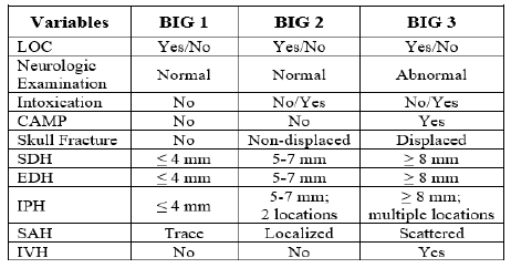
The Brain Injury  Guidelines (BIG) were developed to allow trauma programs to stratify head injuries in such a way as to better utilize resources such as hospital beds, CT scanning, and neurosurgical consultation. Injuries are stratified into three BIG categories, and management is based on it. Here is the stratification algorithm:

And here is the management algorithm based on the stratification above:

The AAST BIG Multi-Institutional Group set about validating this system to ensure that it was accurate and safe. They identified adult patients from nine high level trauma centers that had a positive initial head CT scan. They looked at the the need for neurosurgical intervention, change in neuro exam, progression on repeat head CT, any visits to the ED after discharge, and readmission for the injury within 30 days.

Here are the factoids:

  • About 2,000 patients were included in the study, with BIG1 = 15%, BIG2 = 15%, and BIG3 = 70% of patients
  • BIG1: no patients worsened, 1% had progression on CT, none required neurosurgical intervention, no readmits or ED visits
  • BIG2: 1% worsened clinically, 7% had progression on CT, none required neurosurgical intervention, no readmits or ED visits
  • All patients who required neurosurgical intervention were BIG3 (20% of patients)

The authors concluded that using the BIG criteria, CT scan use and neurosurgical consultation would have been decreased by 29%.

Bottom line: This is an exciting abstract! BIG has been around for awhile, and some centers have already started using it for planning the management of their TBI patients. This study provides some validation that the system works and keeps patients safe while being respectful of resource utilization. 

My only criticism is that the number of patients in the BIG1 and BIG2 categories is low (about 600 combined). Thus, our experience in these groups remains somewhat limited. However, the study is very promising, and more centers should consider adopting BIG to help them refine their management of TBI patients.

Reference: VALIDATING THE BRAIN INJURY GUIDELINES (BIG): RESULTS OF AN AAST PROSPECTIVE MULTI-INSTITUTIONAL TRIAL. AAST 2021, Oral abstract #25.

Antihypertensive Treatment In Acute TBI

Yes, we know high blood pressure can be bad. Over the long term, it can accelerate atherosclerotic heart disease and pound away at the kidneys and brain. And when it is acutely elevated to critical levels, it can lead to stroke.

But is it always bad in trauma? Trauma hurts like hell, so it’s no wonder than many of our patients (not suffering blood loss of course) are hypertensive.  But how often have you seen this scenario occur:

An elderly patient fell from standing, striking her head. She is brought to your ED by ground EMS. She has a GCS of 8 (E1 V3 M4) with a BP of 200/130 and pulse of 56.  This meets your trauma activation criteria and the team assembles to meet the patient.

As you move her onto the bed, one of your colleagues calls out for some nicardipine to control the pressure. Is this a wise move? Remember the First Law of Trauma:

Any anomaly in your trauma patient is due to trauma, no matter how unlikely it may seem.

What else can cause hypertension and bradycardia in your trauma patient? In this case, certainly a subdural or epidural hematoma.

And why is that happening? Because the intracranial pressure is elevated from the space-occupying lesion. Remember the formula for cerebral perfusion pressure (CPP):

CPP = MAP – ICP

Where MAP = mean arterial pressure and ICP = intracranial pressure.  Normally the MAP is around 90 torr and ICP is about 10 torr. Thus, the normal CPP is approximately 80. The range is 60  to well over 100 torr, and flow autoregulation keeps brain perfusion constant over this range.

But let’s say that we are psychic and know the ICP of our patient to be 60 because of a large subdural hematoma. Her current CPP is 150 – 60 or about 90 torr. What happens if we start her on a nicardipine drip or some other antihypertensive medication? We can certainly normalize the blood pressure to 120/80. But now her CPP drops to 90  – 60 = 30 torr!

Congratulations, you have just shut down circulation to her brain!

Bottom line: Think first before calling for antihypertensive medications in patients who may have increased intracranial pressure. You may be sabotaging the only mechanism protecting their brain while you are calling your neurosurgeon for help. Your top priority is to get them to the CT scanner while permitting that pressure. If it turns out that there is no evidence for pathology that would lead to increased ICP, then turn to the antihypertensive agents to help protect against stroke.