Tag Archives: TBI

Diffuse Axonal Injury (DAI)

Trauma professionals tend to focus on the two extremes of TBI: mild concussive injury because we see so much of it, and very severe injury that we have to work so hard to keep the patient alive. Today I’ll write about the one in the middle, diffuse axonal injury (DAI). People don’t talk about it nearly as much, and it seems kind of mysterious.

DAI is also known as a shear injury, because that’s what we think happens to the brain at the time of injury. Officially, it is diagnosed when a patient remains in a coma for more than 6 hours without a demonstrable mass lesion from bleeding seen on CT. It is seen in about 15% of trauma ICU patients with head injury. Essentially the substance of the brain moves around enough to disrupt a critical mass of axonal connections that results in prolonged unconsciousness. It then takes time to try to rebuild those connections and restore consciousness and some degree of cognition. Mechanisms which result in sudden acceleration or deceleration of the brain may cause this condition, and rotational forces which spin the head suddenly seem to be even worse.

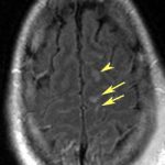
CT scan of the head frequently shows no unusual findings. On occasion, small punctate hemorrhages may be seen. These are generally bad prognostic signs, because CT is so much less sensitive in showing these compared to MRI. Here are some key points about DAI:

  • If the head CT is negative,and all recreational drugs have worn off and the patient still doesn’t wake up, DAI is likely.
  • MRI can confirm the diagnosis, but is not good for giving a prognosis
  • Slow recovery of consciousness or failure to recover correlates with death
  • Hyperglycemia and the presence of a subdural also are highly correlated with mortality

Bottom line: The diagnosis of DAI can generally be made clinically with the assistance of head CT. MRI is not very useful, unless it is needed to confirm the diagnosis. It does not predict speed or degree of recovery so is otherwise not very useful. Supportive care, avoidance of complications and early therapy and rehab are the best treatments we have to offer.

Reference: Diffuse axonal injury in patients with head injuries: an epidemiologic and prognosis study of 124 cases. J Trauma 71(4):838-846, 2011.

Cognitive Rest??

One of the more commonplace recommendations for recovery from mild traumatic brain injury (TBI) is “cognitive rest.” Sports medicine professionals recommend it, physiatrists recommend it, and trauma professionals talk about it.

First, what is it, exactly? I’ve seen a number of descriptions, and they vary quite a bit. The main concept is to avoid all activities that involve mental exertion. This includes using a computer, watching TV, talking on a cell phone, reading, playing video games, and listening to loud music. Huh?

What good does this allegedly do? Most articles that I’ve read theorize that cognitive activity somehow increases the metabolic activity of the brain and that this is bad. One of the more interesting papers I read (from 2010!) says it best: “It is now well-accepted that excessive neurometabolic activity can interfere with recovery from a concussion and that physical rest is needed.”

Read carefully. Well-accepted. The paper cites unpublished data on children by one of the authors, 2 meta-analyses and 2 consensus opinions. In other words, no data at all. Yet somehow the concept has caught on.

First of all, I don’t think it’s possible for most people to realistically practice cognitive rest. Who knows if there is really any difference in metabolism and energy use by the brain if you are engaging in any of the banned activities above? And let’s go to the other extreme: if one lies quietly in bed meditating, shouldn’t this be the ultimate cognitive rest? Yet fMRI and PET studies suggest (also limited data) that cerebral flow in specific areas of the brain increases during this state.

Maybe a modest increase in activity is good. Physical activity (within limits) has been shown to be very beneficial to physical and psychological well being time and time again. And the only paper I could find on the topic with respect to TBI showed that randomization to bedrest vs normal physical activity had no difference in post-concussive syndrome incidence or severity. However, the active group recovered with significantly less dizziness.

Bottom line: There is no data to support the concept of cognitive rest. Any type of activity, either mental or physical, can cause fatigue in a variable amount of time in people with mild TBI. It is probably best to interpret this as a signal to take it easy and recover for a while before exerting oneself again. But so far there is no objective data to show that cognitive activity either helps or hinders recovery.

References:

  • Cognitive rest: the often neglected aspect of concussion management. Athletic Therapy Today, March 2010, pg 1-3.
  • Effectiveness of bed rest after mild traumatic brain injury: a randomised trial of no versus six days of bed rest. J Neurol Neurosurg Psychiatry 73:167-172, 2002.

Technology: Real Time Cerebral Blood Flow Monitoring For TBI

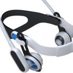
Here’s a new toy that has recently received some funding from the US military. It allows real-time monitoring of cerebral blood flow. It may help identify flow problems from elevated intracranial pressure (ICP) or vasospasm early on, allowing prompt initiation of appropriate therapies to increase blood flow.

This device uses an array of ultrasound beams and locks onto the middle cerebral artery. It then continuously monitors blood flow and displays the result in real time. I predict that there will be a learning curve with this one, similar to near infrared monitoring of tissue perfusion. What’s a normal baseline? What kind of variation is considered “normal?” We’ll have to answer these questions before this tool is ready for prime time. Ultimately, it may allow noninvasive monitoring of ICP in the intensive care unit.

Credit: Physiosonics, Bellevue, WA.

How To Manage TBI In Patients On Warfarin

We all know that the combination of traumatic brain injury (TBI) and warfarin can be dangerous. Here at Regions, we developed a reversal protocol a few years ago. However, we found that just having a list of preferred “antidotes” to give was not enough. The time factor is very important, and we found that we needed to ensure prompt use of these medications when indicated.

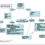
So we added features that ensured timely response and reversal. You can download the protocol by clicking the image above or the link at the bottom of this post.

First, we recognized that any patient with a known or suspected TBI who was taking warfarin was at risk. If the initial GCS was <14, then a full trauma team activation is called. This gives the patient priority lab processing and immediate access to the CT scan. In addition, 2 units of thawed plasma are administered while in the resuscitation room. If the head CT is negative, plasma is stopped.

For patients with a GCS of 14 or 15, a “Code RED” is called, ensuring that an ED physician sees the patient immediately. A point of care INR is drawn and the patient is sent for stat head CT. If the head CT is negative with INR>2.5, the patient is admitted for observation and a repeat head CT is obtained 12 hours later. We have seen patients develop delayed hemorrhage when they have high INR.

We apply a restrictive set of criteria to determine if a patient may go home from the ED, which causes us to admit most for observation. And if they do have a positive CT, we use the algoritm listed below for comprehensive management and reversal.

Bottom line: Patients with any head trauma and an elevated INR are a walking time bomb. They need prompt assessment and reversal of their anticoagulation if indicated. Feel free to share your protocols here as well by posting a comment.

Download the full protocol; click here.

Related post:

Another Anticoagulant To Watch Out For

In May, I wrote about a new direct thrombin inhibitor named dabigatran (Pradaxa). This drug appears beneficial for patients who need ongoing anticoagulation without the hassle of blood testing to check drug levels. The danger for trauma patients is that there is no antidote or rapid reversal possible. This means that significant traumatic bleeding, particularly in and around the brain, cannot be stopped! At Regions Hospital, we have seen a few patients on this drug, but luckily they have not had bleeding from trauma.

Late last month, Bristol-Myers Squibb and Pfizer announced that a new drug has shown very favorable results in preventing strokes in patients with atrial fibrillation (apixaban, Eliquis). Indeed, it cut the relatively low risk of stroke in half, compared to warfarin. It also had about a third fewer bleeding complications. It looks like it may also give dabigatran a run for its money.

This drug is a Factor Xa inhibitor, and also has no antidote other than time. There is some evidence that activated charcoal given orally within 3 hours of apixaban dosing may be somewhat helpful in reducing blood concentrations.

Trauma professionals need to be on the lookout for patients who use this drug. Any trauma patient who admits to being on a “blood thinner” needs to be questioned carefully to determine which one it is. If it is one of the newer drugs without an antidote, they need to be monitored continuously for signs of bleeding (read: ICU), especially if they have experienced head trauma.

Bottom line: Be on the lookout for these drugs. If any patients who have fallen are taking this drug (elderly, frequently intoxicated, etc.), contact their primary physician so that the risks vs benefits of continuing it can be considered.

Related posts


References