Tag Archives: TBI

Resident Work Hour Restrictions And Neurotrauma Complications

In the US, resident work hour restrictions went into effect in 2003, limiting the total number of hours worked per week and the number of consecutive hours without a break. The idea was that fatigue caused errors, which translates into patient complications or worse. Has this panned out? A number of previous publications have found no change; only a few have shown some benefit.

Researchers at Massachusetts General Hospital decided to apply the acid test to this theory. They selected a group of patients who were critically ill and challenging to care for, taken care of by a group of residents who had long work hours and were involved in long operative cases. The AHRQ National Inpatient Sampling Database was studied, comparing the outcomes of neurotrauma patients before and after work hours were initiated and in teaching and non-teaching centers.

A huge number of records were analyzed (40,000 before work hours restrictions, 67,000 after). The findings were intriguing:

  • The overall complication rate was the same before and after restrictions (1.2%)
  • The complication rate was 25% higher in teaching hospitals after restrictions took effect. It appears that this also correlated with higher hospital charges after restrictions.
  • Logistic regression was used to figure out whether this difference was from duty hours or just from the involvement of residents in care. Only duty hours were significant in this analysis.
  • If injury severity was included in the analysis, there were no differences in complications at all
  • There were no differences in mortality rates between any of the groups

Bottom line: Yes, fatigue is bad (see my previous posts below). But here is another (correlation) study that doesn’t bear out the original reasons to restrict resident work hours. In actuality, complications and charges increased after the restrictions went into effect. It is possible that the checks and balances in the system were effective in protecting patients from adverse outcomes. Could the changes in this study be due to staffing changes to meet the restrictions, which results in chronic understaffing which undercuts those checks and balances? Studies of this type can’t tell us that. And unfortunately, restrictions in the US are not going to go away, they’ll probably get worse.

Related posts:

Reference: Higher Complications and No Improvement in Mortality in the ACGME Resident Duty-Hour Restriction Era: An Analysis of More Than 107?000 Neurosurgical Trauma Patients in the Nationwide Inpatient Sample Database. Neurosurgery 70(6):1369-1382, 2012.

Intracranial Hypertension In Pediatric Head Trauma

This 44 minute video is a good introduction to pediatric head trauma and intracranial hypertension. It covers physiology, diagnosis, as well as management using medications, position, decompression and hypothermia.

Presented at Multidisciplinary Trauma Conference at Regions Hospital on May 3, 2012 by Debbie Song MD, a pediatric neurosurgeon.

When To Give Mannitol

Patients with severe head injury need all the help they can get. Mannitol is one tool that is time-tested and cheap. But how do you decide who gets it and when?

Mannitol is a powerful osmotic diuretic that pulls extracellular water from everywhere, including the brain. By reducing the size of the brain overall, it drops pressure inside the skull (ICP) somewhat.

Mannitol can be used anytime during the acute phase of trauma care for three indications in patients with head trauma:

  • Focal neurologic deficit. This is due to transtorial herniation, and may manifest clinically as unilateral pupil dilation or hemiparesis. It may also be seen on CT scan.
  • Progressive neurologic deterioration. This is typical of rising ICP and can be diagnosed when your previously talking patient becomes lethargic.
  • Clinical evidence of high ICP. This is the Cushing response (hypertension with bradycardia). Do not treat this hypertension with other meds, it is a brain protective mechanism!

The literature does not have any good studies that show effectiveness or survival benefit. However, most trauma professionals have seen the dramatic improvement in neurologic status that can occur after early administration.

Bottom line: Mannitol is cheap and it works! Consider it early if any of the three indications above are seen. And don’t forget to put a urinary catheter in immediately because the diuresis that it causes is impressive. And no studies thus far have been able to prove that hypertonic saline is any better or worse than mannitol.

New Technology: Help Brain Injured Patients To Talk

It is can be extremely difficult to communicate with some brain injured patients. Many have global damage that precludes the processing necessary to formulate thoughts. However, some may be able to think but can’t effectively make themselves understood. Patients with the “locked in” syndrome are a perfect example.

A company called NeuroVigil has developed technology and data analysis techniques for extracting a wealth of information from a single-channel EEG. The iBrain system uses two sensors that do not require being stuck to the head with adhesive. A simple elastic band can hold them in place. 

Last year, the company fitted the device on Stephen Hawking to begin testing and training the system to assist with his communication efforts. Currently, Hawking uses an IR sensor that detects twitches in his cheek. These are painstakingly translated into letters and then words that are spoken by a computer. The iBrain system is being trained to recognize words via his EEG patterns and should speed up his communication with the outside world.

If this technology pans out, it may be used to communicate with moderate to severely injured TBI patients who have expressive language problems. It could also be used to test for and communicate with patients who are “locked in.”

The video was recorded at TEDMED 2009. Much of the key information is presented beginning at 10:10 into the video.

I have no financial interest in NeuroVigil

Diffuse Axonal Injury (DAI)

Trauma professionals tend to focus on the two extremes of TBI: mild concussive injury because we see so much of it, and very severe injury that we have to work so hard to keep the patient alive. Today I’ll write about the one in the middle, diffuse axonal injury (DAI). People don’t talk about it nearly as much, and it seems kind of mysterious.

DAI is also known as a shear injury, because that’s what we think happens to the brain at the time of injury. Officially, it is diagnosed when a patient remains in a coma for more than 6 hours without a demonstrable mass lesion from bleeding seen on CT. It is seen in about 15% of trauma ICU patients with head injury. Essentially the substance of the brain moves around enough to disrupt a critical mass of axonal connections that results in prolonged unconsciousness. It then takes time to try to rebuild those connections and restore consciousness and some degree of cognition. Mechanisms which result in sudden acceleration or deceleration of the brain may cause this condition, and rotational forces which spin the head suddenly seem to be even worse.

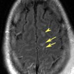
CT scan of the head frequently shows no unusual findings. On occasion, small punctate hemorrhages may be seen. These are generally bad prognostic signs, because CT is so much less sensitive in showing these compared to MRI. Here are some key points about DAI:

  • If the head CT is negative,and all recreational drugs have worn off and the patient still doesn’t wake up, DAI is likely.
  • MRI can confirm the diagnosis, but is not good for giving a prognosis
  • Slow recovery of consciousness or failure to recover correlates with death
  • Hyperglycemia and the presence of a subdural also are highly correlated with mortality

Bottom line: The diagnosis of DAI can generally be made clinically with the assistance of head CT. MRI is not very useful, unless it is needed to confirm the diagnosis. It does not predict speed or degree of recovery so is otherwise not very useful. Supportive care, avoidance of complications and early therapy and rehab are the best treatments we have to offer.

Reference: Diffuse axonal injury in patients with head injuries: an epidemiologic and prognosis study of 124 cases. J Trauma 71(4):838-846, 2011.