Tag Archives: imaging

How Much Radiation Exposure In Imaging Studies?

Everyone knows that CT scans deliver more radiation than conventional x-ray. But how much does each test really deliver? And how significant is that?

Let me try to put it all into perspective. First, how much radiation are we exposed to just living outside the hospital? Background radiation is everywhere. It consists of radioactive gases (argon) in the air we breathe, radiation from the rocks and other things around us, and cosmic rays blasting through us from space.

In the United States, the average background radiation each of us is exposed to is about 3.1 milliSieverts (mSv). I’ve compiled a table to show the approximate dose delivered by some of the common radiographic studies ordered by trauma professionals. And to keep it real, I’ve calculated how much extra background radiation we would have to absorb, in units of time, to have an equivalent exposure.

Read and enjoy! Remember, doses may vary by scanner, settings, and dose reduction measures used.

Test Dose (mSv) Equivalent background
radiation
Chest x-ray 0.1 10 days
Pelvis x-ray 0.1 10 days
CT head 2 8 months
CT cervical spine 3 1 year
Plain c-spine 0.2 3 weeks
CT chest 7 2 years
CT abdomen/pelvis 10 3 years
CT T&L spine 7 2 years
Plain T&L spine 3 1 year
Millimeter wave
scanner (that hands
in the air TSA thing at
the airport)
0.0001 15 minutes
Scatter from a chest
x-ray in trauma bay
when standing one 
meter from the
patient
0.0002 45 minutes
Scatter from a chest
x-ray in trauma bay
when standing three 
meters from the
patient
0.000022 6 minutes

EAST 2019 #10: Incidental Findings In Trauma Imaging

Every major trauma patient undergoes some type of radiographic imaging during their initial evaluation. On occasion, some incidental finding unrelated to trauma shows up unexpectedly. These incidentalomas add several additional layers of complexity to the evaluation process.

What does the finding mean? Is it important? How do I tell the patient? Their primary care provider? When? Many times, these findings have little clinical significance. But on occasion, they can be life changing, such as the incidental renal cell carcinoma.

The group at University of Tennessee – Knoxville reviewed one year of incidental findings in trauma evaluations at their Level I trauma center. They specifically looked at diagnoses with malignant potential, and how findings were disclosed to the patient.

Here are the factoids:

  • Over 6000 patients were reviewed, and 22% had 1222 incidental findings (that’s 2 per patient!)
  • The findings were noted in males about 2/3 of the time
  • 59% of of incidentalomas were in the chest, and 16% in the abdomen
  • The most common findings were lung nodule (209), hernia (112), and renal cyst (103)
  • Only 60% of patients were informed prior to discharge (!)
  • Trauma registry abstraction resulted in an additional 20% of patients informed of the finding
  • 58 patients could not be located, and in 43 patients there was no documented attempt to contact them
  • An additional 100 registry charts that did not contain incidental findings were re-abstracted and searched for incidental findings. Nearly one third contained incidental findings!
  • If the incidental finding was noted in the radiology report summary, 78% of patients were informed. But when it was buried in the body of the report, only 22% were disclosed.

Here are some questions for the authors and presenter to consider in advance to help them prepare for audience questions:

  • The majority of the incidental findings were in the chest and abdomen. What and where were the rest?
  • What would you recommend for achieving optimal disclosure based on your results? It appears that 20% or so of patients never learned of the finding.
  • What should we do about our registry data? Should we force our registrars to comb all reports for possible incidental findings? Given that one fifth of patients have them (or more) that seems like a lot of work!
  • How has your work changed your practice at UT Knoxville?

This is a fascinating paper, and gives me some ideas for upcoming blog posts! I will definitely be in the audience for this presentation.

Reference:  A novel use of the trauma registry: incidental findings in the trauma patient. EAST 2019, Quick Shot Paper #13.

Pan Scanning for Elderly Falls?

The last abstract for the Clinical Congress of the American College of Surgeons that I will review deals with doing a so-called “pan-scan” for ground level falls. Apparently, patients at this center have been pan-scanned for years, and they wanted to determine if it was appropriate.

This was a retrospective trauma registry review of 9 years worth of ground level falls. Patients were divided into young (18-54 years) and old (55+ years) groups. They were included in the study if they received a pan-scan.

Here are the factoids:

  • Hospital admission rates (95%) and ICU admission rates (48%) were the same for young and old
  • ISS was a little higher in the older group (9 vs 12)
  • Here are the incidence and type of injuries detected:
Young (n=328) Old (n=257)
TBI 35% 40%
C-spine 2% 2%
Blunt Cereb-vasc inj * 20% 31%
Pneumothorax 14% 15%
Abdominal injury 4% 2%
Mortality * 3% 11%

 * = statistically significant

Bottom line: There is an ongoing argument, still, regarding pan-scan vs selective scanning. The pan-scanners argue that the increased risk (much of which is delayed or intangible) is worth the extra information. This study shows that the authors did not find much difference in injury diagnosis in young vs elderly patients, with the exception of blunt cerebrovascular injury.

Most elderly patients who fall sustain injuries to the head, spine (all of it), extremities and hips. The torso is largely spared, with the exception of ribs. In my opinion, chest CT is only for identification of aortic injury, which just can’t happen from falling over. Or even down stairs. And solid organ injury is also rare in this group.

Although the future risk from radiation in an elderly patient is probably low, the risk from the IV contrast needed to see the aorta or solid organs is significant in this group. And keep in mind the dangers of screening for a low probability diagnosis. You may find something that prompts invasive and potentially more dangerous investigations of something that may never have caused a problem!

I recommend selective scanning of the head and cervical spine (if not clinically clearable), and selective conventional imaging of any other suspicious areas. If additional detail of the thoracic and/or lumbar spine are needed, specific spine CT imaging should be used without contrast.

Related posts:

Reference: Pan-scanning for ground level falls in the elderly: really? ACS Surgical Forum, trauma abstracts, 2016.

Radiographic Image Sharing Systems

There are generally three ways to share radiographic images with your upstream trauma center:

  • Hard copy. These days, that usually means a CD. Nearly all PACS systems (picture archiving and communications systems) can write CDs that can accompany your patient. Advantage: super cheap. Possible downsides: the CD may be corrupted and not openable, the software on the disk cannot be installed or will not run at the receiving hospital, and finally it can just be forgotten in the rush to get the patient out of the ED.
  • PACS system connections. These are software links that enable one hospital’s PACS software to communicate with another’s. They must be established in advance, and generally require some expertise from the hospitals’ IT departments. Images can be pushed from one system to another. Advantages: once set up, it is very inexpensive to maintain, and images can be viewed prior to patient arrival at the receiving hospital. Possible downside: Al-though the interchange format is standardized, every once in a while the systems just can’t communicate.
  • Web-based image sharing system. This consists of a web server-based software application available via the internet that allows subscribing hospitals to sign on and share images. Referring hospitals can upload images from their PACS systems for free, and the receiving hospital can view the images and/or download into their own system. Advantage: these products are simple to set up, and easy to use after just a little training. Compatibility is very high, and the services are continually working to ensure it. Downside: expensive. Depending on specifics, the annual subscription may be up to $100K per year, and is generally footed by the receiving trauma center.

Is a web-based solution worth it? MetroHealth in Cleveland looked at this over five years ago, and published their results in 2015. They looked at their experience pre- and post-implementation and found the following:

  • Three years of transfer data prior to the web system implementation was compared to one year of experience after
  • CT imaging decreased at both referring and receiving hospitals across the study period
  • Repeat scan rate decreased from 38% to 28%. Repeat head scans were the major driver at 21%.
  • Cost of reimaging dropped from about $1000 per patient to $600

Bottom line: As a referring hospital, it is your responsibility to ensure that the (hopefully) few images you obtain make it to the upstream trauma center. Although hard copy (CD) is the cheapest, it is also the least reliable. Work with your radiology and IT departments to determine which electronic solution is best for you. Some states and regional trauma systems help subsidize or provide a web-based solution for their member hospitals.

Reference: Implementation of an image sharing system significantly reduced repeat computed tomographic imaging in a regional trauma system. J Trauma 80(1):51-56, 2016.

Part 2: Metal Splints – Can You CT Scan Through Them?

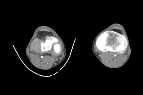
In my last post, I debunked the myth that using a pre-formed aluminum splint significantly degrades the quality of standard x-rays. But what about a study that provides much more detail, such as CT scan?

CT scan techs have told me that there would be too much artifact using any kind of metal splint. And typically, when imaging an extremity with CT, we are looking at vascular runoff. The vessels are small, and high image quality is extremely important. If the images are bad, then we risk having to give the patient another dose of both radiation and contrast.

As you know, my mantra is question everything! So i scouted around and found some images to share using one of these splints. Look closely for the intimal flap in the image below:

Can’t see it? That’s because it isn’t there! But you certainly could if it were!

Bottom line: A perforated aluminum splint causes absolutely no artifact or image degradation. Do not cause additional injury by removing it prior to imaging, either CT or conventional x-ray. Although your friendly techs, radiologists, and orthopedic surgeons may moan, it won’t hurt their ability to make decisions on the images.