Tag Archives: fracture

Best Of EAST 2023 #9: CT Of The Lumbar Spine

It is becoming clearer and clearer that patients with suspicion for fractures of the thoracic (T) and lumbar (L) spine should be imaged only with CT scan. Conventional imaging just doesn’t have enough sensitivity, even in younger patients with healthy bones. But when we obtain CT of the T&L spines there is a choice: just look at the axial / helical slices, or have the computer reconstruct additional images in the sagittal and coronal planes. The belief is that this multiplanar imaging will assist in finding subtle fractures that might not be seen on axial views.

The group at Rutgers in New Jersey tried to determine if adding the reconstructions amounted to overkill. They performed a retrospective review of patients at their Level I center over a six-year period. They focused on studies performed in patients who had T and/or L fractures who also had both CT of the chest, abdomen, and pelvis (CAP) and thoracic and lumbar reconstructions. Additional data were obtained from a review of the medical record and trauma registry.

Here are the factoids:

  • A total of 494 patients had both CAP and reconstructions
  • There were 1254 fractures seen on CAP, and an additional 129 fractures seen with recons (total of 1394)
  • The majority of additional injuries not detected on CT CAP were transverse process fractures
  • The number of other fracture patterns not seen on CT CAP were statistically “not significant”
  • However, these numerically “not significant” fractures included 51 vertebral body fractures, 6 burst fractures, 3 facet fractures, and 2 pedicle fractures
  • No unstable fractures were missed on CT CAP
  • More MRIs were performed in the patients who had recons, there were more spine consultations, and 11% underwent operative fixation vs. 2% for CTA only (!!)

The authors concluded that CT CAP alone was sufficient to identify clinically significant thoracic and lumbar fractures. They also stated that clinically insignificant injuries identified with reconstructions were more likely to undergo MRI and use excess resources. They urged us to be selective with the use of T&L reformats.

Bottom line: Wow! I have a lot of questions about this abstract! And I really disagree with the findings.

You studied fewer than 500 patients with T or L spine fractures over a six year period. This is only about 80 per year, which seems very low. This suggests that many, many patients were being scanned without recons to start with. How did patients get selected out to get recons? Were there specific criteria? I worry that this could add some bias to your study.

The number of fractures seen only on the recon views besides transverse process fractures were deemed “statistically insignificant.” However, looking at the list of them (see bullet point 5 above) they don’t look clinically insignificant. It’s no wonder that recons resulted in more consults, MRI scans, and spine operations!

I worry that your conclusion is telling us to stop looking for fractures so we won’t use so many additional resources. But their use may be in the best interest of the patients!

Here are my questions and comments for the presenter/authors:

  • Why did you decide to do this study? I didn’t realize that not doing the recons was a thing in major blunt trauma. Was there some concern that resources were being wasted? Was there an additional cost for the reconstructions?
  • How many patients only received CT CAP? The greater the number of these, the higher the probability that some non-random selection process is going on that might bias your findings.
  • How did you get separate reports for the non-reconstructed images? Did you have new reads by separate radiologists? Typically, the report contains the impression for the entire study. It would be unusual for the radiologist to comment on the non-recon images, then add additional findings from just the reconstructions.
  • Doesn’t the increased numbers of spine consults, MRIs, and operative procedures in the patients with reconstructions imply that these otherwise occult fractures needed clinically important additional attention? 

I worry that readers of this abstract might take away the wrong message. Unless there is some additional compelling data presented, this study is certainly not enough to make me change my practice!

Reference: UTILITY OF CT THORACOLUMBAR SPINAL RECONSTRUCTION IMAGING IN BLUNT TRAUMA. EAST 2023 Podium paper #20.

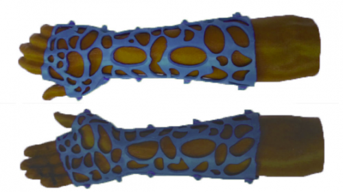
Fracture Care Of The Future: Traditional Casts vs 3D-Printed Braces

I’ve been fascinated by 3D printing for at least a decade.  Here are some examples from previous posts:

Unfortunately, practical applications have been relatively limited in the field of trauma.  But a lot has been going on in the background. The trauma research group at Erasmus Medical Center in Rotterdam recently published a systematic review on very practical work using 3D printing to produce casts and splints.

Sounds like a very mundane problem to through high tech at, right? But for those of you who look after patients with fractures that have been casted, you know the problems that can arise. Casts can be too tight. They can be ill-fitting. The patient may have soft tissue injuries that require windows cut into the side of the cast. Additional technology such as electrical stimulators may be indicated to enhance healing.

The old-fashioned way of creating a plaster or fiberglass cast seems crude. It is shaped by hand using skill and a fair amount of guesswork. If it’s just a bit too tight, serious complications may occur. If windows are not cut properly, it can destabilize the entire cast.

The Rotterdam trauma research group performed a systematic review of 12 papers that have been published on the topic of 3D-printed casts used in the treatment of forearm fractures. The authors found that most currently use a technique called fused deposition modeling with a polylactic acid substrate.

Instead of relying on subjective skill and luck to shape the brace, the uninjured forearm is scanned with a 3D scanner. The data is fed to a computer aided design (CAD) workstation and a mirror image is created and further refined. Special features such as soft tissue windows or entry points for bone stimulators can be designed into the brace at that time. Because the strength of polycarbonate exceeds that of plaster and fiberglass, it is possible to create a design with a great deal of open area so the underlying skin can be monitored. And allowances can be made for areas with swelling not present on the control extremity.

The data is then fed to a 3D printer to actually create the cast. Here’s an example:

This design is stronger that a traditional cast, is cool and comfortable, and avoids problems with hidden tissue injury or unrecognized foreign objects dropping into the cast creating major problems.

The use of 3D-printed casts and braces is relatively new and is used in only a few centers. For this reason, we do not have enough numbers to show that it is equivalent to traditional casting. Yet. But as the price continues to drop and use becomes more widespread, it’s only a matter of time before you start seeing these items in your own trauma center.

Reference: Personalize d 3D-printed forearm braces as an alternative for a traditional plaster cast or splint; A systematic review. Injury, in press, July 29, 2022. https://doi.org/10.1016/j.injury.2022.07.020

The Impact Of NSAIDs On Fracture Healing

In my last post I discussed some of the basic effects of NSAIDs on bone healing. Now let’s see if theory applies to practice.

In 2003, several papers brought to light possible interactions between these drugs and fracture healing. Specifically, there were questions about these drugs interfering with the healing process and of increasing the number of delayed unions or nonunions. But once again, how convincing were these papers, really?

It would seem to make sense that NSAIDs could interfere with bone healing. The healing process relies heavily on the regulation of osteoblast and osteoclast function, which itself is regulated by prostaglandins. Since prostaglandins are synthesized by the COX enzymes, COX inhibitors like the NSAIDs should have the potential to impair this process. Indeed, animal studies in rats and rabbits seem to bear this out.

But as we have seen before, good animal studies don’t always translate well into human experience. Although a study from 2005 suggested that NSAID administration in older patients within 90 days of injury had a higher incidence of fracture nonunion, the study design was not a very good one. It was equally likely that patients who required these drugs in this age group may have been at higher risk for nonunion in the first place.

A meta-analysis of human studies was performed in 2011. Out of 558 potential studies, only 5 met criteria review. (This is yet another reminder of the sheer amount of sub-par research out there.) The authors found that short-term use (< 14 days) of normal dose NSAIDS was not associated with non-union. High doses of ketorolac (> 120mg/day) and diclofenac sodium (> 300mg total) did have an association. But remember, this does not show causation. There are many other factors that can impede healing (smoking, diabetes, etc).

A study from 2016 examined the effect of ketorolac administration on fracture healing in patients undergoing repairs of femoral and tibial fractures. It did not find an association between non-union and ketorolac, but did find one with smoking. Unfortunately, the study was small (85 patients given ketorolac, 243 controls without it). It probably does not have the statistical power to detect any difference with the NSAID. A power analysis was not provided in the methods section.

Bottom line: Once again, the animal data is clear and the human data less so. Although there are theoretical concerns about NSAID use and fracture healing, there is still not enough solid risk:benefit information to abandon short-term NSAID use in patients who really need them. NSAIDs can and should be prescribed in patients with short-term needs and simple fractures, and consider COX-1 specific drugs like ketorolac while your patient is in the hospital. And we do have some evidence that high-dose NSAIDs may have some impact, so stick to the usual doses for just as long as they are needed for pain management.

References:

  1. Effects of nonsteroidal anti-inflammatory drugs on bone formation and soft-tissue healing. J AM Acad Orthop Surg 12:139-43, 2004.
  2. Effect of COX-2 on fracture-healing in the rat femur. J Bone Joint Surg Am 86:116-123, 2004.
  3. Effects of perioperative anti-inflammatory and immunomodulating therapy on surgical wound healing. Pharmacotherapy 25:1566-1591, 2005.
  4. Pharmacological agents and impairment of fracture healing: what is the evidence? Injury 39:384-394, 2008.
  5. High dose nonsteroidal anti-inflammatory drugs compromise spinal fusion. Can J Anaesth 52:506-512, 2005.
  6. Nonsteroidal Anti-Inflammatory Drugs and Bone-Healing: A Systematic Review of Research Quality. JBJS Rev 4(3), 2016.
  7. High-dose ketorolac affects adult spinal fusion. Spine 36(7):E461-E468, 2011.
  8. Ketorolac administered in the recovery room for acute pain management does not affect healing rates of femoral and tibial fractures. J Orthop Trauma 30(9):479-482, 2016.

Salmon Calcitonin After Spine Fractures?

In my last post, I reviewed some data on the effectiveness of starting Vitamin D supplements after a patient sustains a fracture. The idea was that they might start building better bone and heal their fractures more readily if they boost their D levels. Unfortunately, this was not shown to be true.

Vitamin D improves bone health by facilitating absorption of calcium from the gut. This is a bit indirect and relies on sufficient intake of calcium and good hormonal regulation that directs osteoblasts to incorporate the mineral into bone. Why not work with those hormones directly to try to increase the amount of calcium that is deposited?

Calcitonin is a peptide hormone that has two major effects on calcium levels: it inhibits osteoclast activity that is breaking down bone and releasing calcium from it, and it inhibits calcium reabsorption in the kidneys which causes more to be excreted in the urine.

Perhaps giving calcitonin after sustaining a fracture might improve healing. Many orthopedic surgeons and neurosurgeons swear by this drug. Unfortunately, there are very few randomized, controlled studies of its use for this indication. A meta-analysis was performed that examined both utility and cost-effectiveness that I found interesting.

Here are the factoids:

  • There was some mild evidence that nasal calcitonin was effective in preventing vertebral fractures
    • One paper showed a benefit when giving 200 IU of intranasal salmon calcitonin daily over a 5 year period
    • But a benefit was not shown if 100 or 400 IU were given (this is weird)
    • A marker of bone turnover showed equal reduction in 200 and 400 IU groups (why isn’t this less in the 200 IU group?)
  • Financial analysis showed that it was only marginally cost effective
  • Current retail pricing is about $125 for a month supply
  • Mild side effects like runny nose and nausea are common
  • Intranasal calcitonin has been shown to reduce pain during healing of vertebral fractures

Bottom line: What does all of this mean? First, salmon calcitonin might decrease the number of future vertebral fractures. I say might because only the 200 IU dose in the study showed this effect. I can see where higher doses might be more effective to a point, but having only the middle dose show up as effective is just odd and makes me worry about the study.

The data does seem compelling that taking this product decreases pain during fracture healing. A meta-analysis of this showed that the effect probably only lasts up to a month. 

And finally, from a cost-effectiveness standpoint for avoiding future fractures, this medication is marginal. Luckily, it is relatively cheap at $125 retail and about $25 with insurance in the US. 

Wrapping it all up, intranasal salmon calcitonin might reduce fracture pain for a month and might decrease the likelihood of future vertebral fractures. However, the data are weak enough that cost-effectiveness is borderline. And there are more effective (and cheaper) analgesics available.

The absolute best way to strengthen bones is to exercise, especially engaging in weight-bearing activities. Not only does this strengthen bones, it also increases overall fitness and health. In general, medications are not the way to go to strengthen bones. It took decades for your patient to become osteoporotic. And while these drugs might improve their bone density slowly, a graduated and supervised exercise regimen is probably the best thing you can do for them.

References:

  • Efficacy of calcitonin for treating acute pain associated with osteoporotic vertebral compression fracture: an updated systematic review. CJEM 2020 May;22(3):359-367.
  • A Randomized Trial of Nasal Spray Salmon Calcitonin in Postmenopausal Women with Established Osteoporosis: the Prevent Recurrence of Osteoporotic Fractures Study. PharmacoEconomics, 2001, Vol.19 (5), p.565-575.
  • A randomized, double-blind, multicenter, placebo-controlled study to evaluate the efficacy and safety of oral salmon calcitonin in the treatment of osteoporosis in postmenopausal women taking calcium and vitamin D. Bone 2016 Oct;91:122-9.

Giving Vitamin D After Fracture: Helpful Or Not?

The role of Vitamin D in fracture healing is well known. So, of course, trauma professionals have tried to promote Vitamin D supplementation to counteract the effects of osteoporosis. A meta-analysis of of 12 papers on the topic relating to hip and other non-vertebral fractures showed that there was roughly a 25% risk reduction for any non-vertebral fractures in patients taking 700-800 U of Vitamin D supplements daily.

Sounds good, right? So what about taking Vitamin D after a fracture occurs? Seems like it should promote healing, right? A large meta-analysis in an orthopedics journal looked at this very question.

Unfortunately, there was a tremendous variability in the interventions, outcomes, and measures of variance. All the authors could do was summarize individual papers, and a true meta-analysis could not be performed.

Here are the factoids:

  •  81 papers made the cut for final review
  • A whopping 70% of the population with fractures had low Vitamin D levels
  • Vitamin D supplementation in hospital and after discharge did increase serum levels
  • Only one study, a meeting abstract which has still not seen the light of day in a journal, suggested a trend toward less malunions following a single loading dose of Vitamin D

Bottom line: Vitamin D is a great idea for people who are known to have, or are at risk for, osteoporosis and fractures. It definitely toughens up the bones and lowers the risk of fracture. However, the utility of giving it after a fall has not been shown. Of the 81 papers reviewed, none showed a significant impact on fracture healing. The only good thing is that Vitamin D supplements are cheap. Giving them may make us think that we are helping our patient heal, but it’s not. 

What about the use of calcitonin for preventing future fractures? Find that in my next post!

References:  

  • What is the role of vitamin D supplementation in acute fracture patients? A systematic review and meta-analysis of the prevalence of hypovitaminosis D and supplementation efficacy. J Orthopaedic Trauma 2016 Feb;30(2):53-63.
  • Fracture prevention with vitamin D supplementation: a meta-analysis of randomized controlled trials. JAMA 293(18):2257-2264, 2005.