Tag Archives: DVT

Pulmonary Embolism and DVT in Trauma

We have long assumed that pulmonary emboli start as clots in the deep veins of the legs (or pelvis), then break off and float into the branches of the pulmonary artery in the lungs. A huge industry has developed around how best to deal with or prevent this problem, including mechanical devices (sequential compression devices), chemical prophylaxis (heparin products), and physical devices (IVC filters).

The really interesting thing is that less than half of patients who are diagnosed with a pulmonary embolism have identifiable clots in their leg veins. In one study, 26 of 200 patients developed DVT and 4 had a PE. However, none of the DVT patients developed an embolism, and none of the embolism patients had a DVT! How can this kind of disparity be explained?

Researchers at the Massachusetts General Hospital retrospectively looked at the correlation between DVT and PE in trauma patients over a 3 year period. DVT was screened for on a weekly basis by duplex venous ultrsonagraphy. PE was diagnoses exclusively using CT scan of the chest, but also included the pelvic and leg veins to look for a source. A total of 247 patients underwent the CT study for PE and were included in the study.

Here are the factoids:

  • Forty six patients had PE (39% central, 61% peripheral pulmonary arterial branches) and 18 had DVT (16 seen on the PE CT and 2 found by duplex)
  • Of the 46 patients with PE, only 15% had DVT
  • All patient groups were similar with respect to injuries, injury severity, sex, anticoagulation and lengths of stay
  • Interestingly, 71% of PE patients with DVT had a central PE, but only 33% of patients without DVT had a central PE.

The authors propose 4 possible explanations for their findings:

  1. The diagnostics tools for detecting DVT are not very good. FALSE: CT evaluation is probably the “gold standard”, since venography has long since been abandoned
  2. Many clots originate in the upper extremities. FALSE: most centers do not detect many DVTs in the arms
  3. Leg clots do not break off to throw a PE, they dislodge cleanly and completely. FALSE: cadaver studies have not shown this to be true
  4. Some clots may form on their own in the pulmonary artery due to endothelial inflammation or other unknown mechanisms. POSSIBLE

An invited critique scrutinizes the study’s use of diagnostics and the lack of hard evidence of clot formation in the lungs.

Bottom line: this is a very intriguing study that questions our assumptions about deep venous thrombosis and pulmonary embolism. More work will be done on this question, and I think the result will be a radical change in our use of anticoagulation and IVC filters over the next 3-5 years.

Reference: Pulmonary embolism and deep venous thrombosis in trauma: are they related? Arch Surg 144(10):928-932, 2009.

How To Predict Venous Thromboembolism In Pediatric Trauma

As with adults a decade ago, the incidence of venous thromboembolism (VTE) in children is now on the rise. Whereas adult VTE occurs in more than 20% of adult trauma patients without appropriate prophylaxis, it is only about 1% in kids, but increasing. There was a big push in the early 2000′s to develop screening criteria and appropriate methods to prevent VTE. But since the incidence in children was so low, there was no impetus to do the same for children.

The group at OHSU in Portland worked with a number of other US trauma centers, and created some logistic regression equations based on a large dataset from the NTDB. The authors developed and tested 5 different models, each more complex than the last. They ultimately selected a model that provided the best fit with the fewest number of variables.

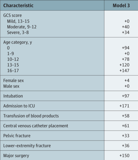
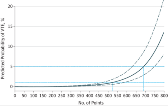
The tool consists of a list of risk factors, each with an assigned point value. The total point value is then identified on a chart of the regression equation, which shows the risk of VTE in percent.

Here are the factors:

Note that the highest risk factors are age >= 13, ICU admission, and major surgery.

And here is the regression chart:

Bottom line: This is a nice tool, and it’s time for some clinical validation. So now all we have to do is figure out how much risk is too much, and determine which prophylactic tools to use at what level. The key to making this clinically usable is to have a readily available “VTE Risk Calculator” available at your fingertips to do the grunt work. Hmm, maybe I’ll chat with the authors and help develop one!

Reference: A Clinical Tool for the Prediction of Venous Thromboembolism in Pediatric Trauma Patients. JAMA Surg 151(1):50-57, 2016.

EAST 2016: How Long Does VTE Risk Last In TBI?

Most trauma patients
are considered to be at some risk for deep venous thrombosis (DVT) and/or
venous thromboembolism (VTE) during their hospital stay. Trauma professionals
go to great lengths to screen for, prophylax against, and treat these problems.
One of the tougher questions is, how long do we need to worry about it? For
fractures, we know that the risk can persist for months. But what about head
injury?

A group at Brigham
and Women’s Hospital did a large database study looking at the VTE risk in adults
who sustained significant head injury, with only minor injuries to other body
regions. They tried to tease out the risk factors using multivariate regression
models.

Here are the
factoids:

  • Patients were only included if their AIS Head
    was >3, and all other AIS were <3
  • Of the over 50,000 patients in the study,
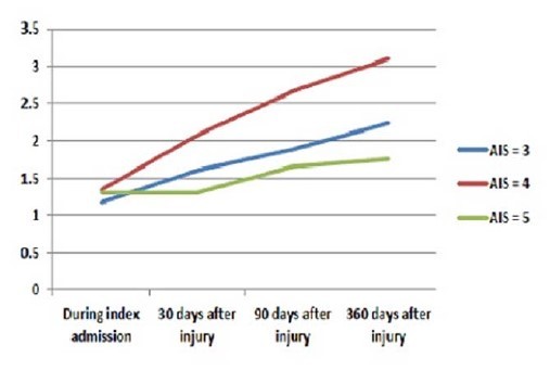
    overall incidence of VTE was 1.3% during the hospital stay, and 2.8% overall
    within 1 year
    of injury
  • Risk factors for VTE after discharge included
    age > 64 (3x), discharge to a skilled nursing facility (3x), and prolonged
    hospital length of stay
    (2x)
image

Incidence of VTE over time

Bottom line: View this paper as a glimpse of a potential unexpected
issue. The risk of VTE persists for quite some time after head injury (and
probably in most other risky injuries like spine and pelvic fractures. The
three risk factors identified seem to identify a group of more seriously
injured patients who do not return to their baseline soon after injury. We may
need to consider a longer period of screening in select patients, but I believe
further work needs to be done to help figure out exactly who they are.

Reference: How long should we fear? Long-term risk of
venous thromboembolism in patients with traumatic brain injury. EAST 2016 Oral
abstract #28.

How Long Are Trauma Patients At Risk For VTE?

Venous thromboembolism (VTE) and its complications are one of the banes of the trauma professional’s existence. Trauma centers have initiated extensive systems of risk assessment, screening, prophylaxis, and treatment of patients at risk for this problem. But typically, much of this management ends at or shortly after discharge from the hospital.

How long do we need to worry? Some trauma programs continue prophylaxis on at-risk patients until they are ambulating well, or for an arbitrary period of time, like one month. But until recently, we’ve had no guidance based on actual numbers. A California study may shed some light on this gray area.

A large dataset from a state of California hospital discharge database was massaged, looking at 6 years of data from patients at the highest risk for VTE (injuries of the pelvis, spine, and spinal cord). The authors looked forward in time after the initial discharge to see if there were any future admissions for VTE and its complications. 

Here are the factoids:

  • Patients with spinal cord injury had the highest risk of VTE, pelvic fractures were mid-range, and vertebral fractures the lowest risk.
  • Occurrence of VTE was associated with a significant risk of mortality, but it was not possible to determine why.
  • In all groups, the risk of VTE remained for the first 3 months after injury, then declined rapidly.
  • VTE risk returned to the level of the general population after about 12 months in patients with pelvic and vertebral injuries.
  • VTE risk in spinal cord injured patients followed a similar curve, but never completely returned to the population baseline.

Bottom line: Obviously, this is not a clinical study. But it’s size and duration is unprecedented and provides valuable information anyway. This information calls into question our existing treatment intervals for prevention of VTE. However, it does not provide real and actionable guidance yet. Additional clinical studies will be needed to parse out the best drugs and duration of treatment.

Related posts:

Reference: Can we ever stop worrying about venous thromboembolism after trauma? J Trauma 78(3):475-481, 2015.

More On DVT In Children

Deep venous thrombosis has been a problem in adult trauma patients for some time. Turns out, it’s a problem in injured children as well although much less common (<1%). However, the subset of kids admitted to the ICU for trauma have a much higher rate if not given prophylaxis (approx. 6%). Most trauma centers have protocols for chemical prophylaxis of adult patients, but not many have similar protocols for children.

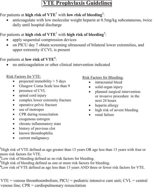
The Medical College of Wisconsin looked at trends prior to and after implementation of a DVT protocol for patients < 19 years old. They used the following protocol to assess risk in patients admitted to the PICU and to determine what type of prophylaxis was warranted:

image

The need for and type of prophylaxis was balanced against the risk for significant bleeding, and this was accounted for in the protocol. The following significant findings were noted:

  • The overall incidence of DVT decreased significantly (65%) after the protocol was introduced, from 5.2% to 1.8%
  • The 1.8% incidence after protocol use is still higher than most other non-trauma pediatric populations 
  • After the protocol was used, all DVT was detected via screening. Suspicion based on clinical findings (edema, pain) only occurred pre-implementation.
  • Use of the protocol did not increase use of anticoagulation, it standardized management in pediatric patients

Bottom line: DVT does occur in injured children, particularly in severely injured ones who require admission to the ICU. Implementation of a regimented system of monitoring and prophylaxis decreases the overall DVT rate and standardizes care in this group of patients. This is another example of how the use of a well thought out protocol can benefit our patients and provide a more uniform way of managing them.

Related posts:

Reference: Effectiveness of clinical guidelines for deep vein thrombosis prophylaxis in reducing the incidence of venous thromboembolism in critically ill children after trauma. J Trauma 72(5):1292-1297, 2012.