Tag Archives: ct scan

More On CT Usage In Unstable Patients

Yes, it is practically dogma that CT should not be used in unstable trauma patients. Either they go directly to the OR, or an attempt to stabilize them is briefly undertaken in the trauma bay. And as you know, I’m not a big believer in dogma. But this one has withstood the test of time. You can see my comments about a previous paper below in the related posts.

But now some authors in Colombia have published a paper that seems to call this idea into question. Could it be true? Read carefully!

This was a small, retrospective review of patients from a large Level I government designated trauma center. They reviewed their experience over a two year period, identifying all hemodynamically unstable patients in the registry. They excluded dead patients, those with isolated head injury, and any who had surgery at an outside hospital prior to transfer.

Here are the factoids:

  • 171 patients were reviewed, and of course they tended to be young males
  • 91 went straight to the OR, and 80 were taken to CT first
  • “Unstable” patients were defined as having SBP < 100 and/or HR > 100
  • Mechanism of injury for the OR group was 95% penetrating, but for the CT group was about 50:50 penetrating/blunt
  • The mean SBP and HR for the “unstable” patients taken to CT were 92 and 110, respectively
  • Mortality was the same for both groups (18% OR vs `13% CT)

Bottom line: The authors concluded that it is permissible to take unstable patients to CT if you don’t spend too much time there based on similar mortality rates. But the problem was that I don’t consider their patients to have been unstable! Mean SBP in their “unstable” group was over 90 torr and the heart rate was only 110! The lowest SBP was only 79. And mortality is too crude of an outcome to rely on. Furthermore, the patients they took to CT tended to have blunt mechanisms, and may not have had ample efforts at resuscitation in the trauma bay first, or may have met criteria to go to CT anyway (see related posts below).

Reference: Computed tomography in hemodynamically unstable severely injured blunt and penetrating trauma patients. J Trauma 80(4):597-603, 2016.

Impact Of Arm Position On Torso CT Scan

CT scan is a valuable tool for initial screening and diagnosis of trauma patients. However, more attention is being paid to radiation exposure and dosing. Besides selecting patients carefully and striving for ALARA radiation dosing (as low as reasonably achievable) by adjusting technique, what else can be done? Obviously, shielding parts of the body that do not need imaging is simple and effective. But what about simply changing body position?

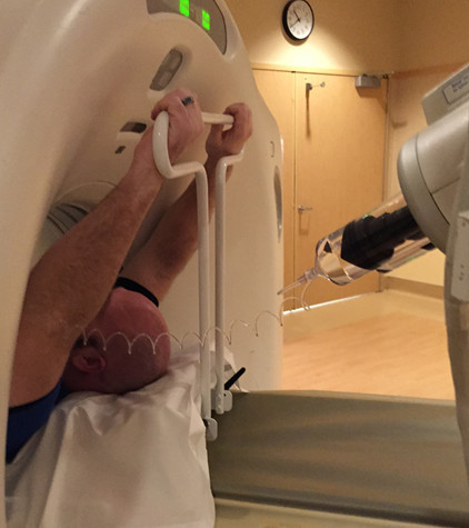
One simple item to consider is arm positioning in torso scanning. There are no consistent recommendations for use in trauma scanning. Patients with arm and shoulder injuries generally keep the affected upper extremity at their side. Radiologists prefer to have the arms up if possible to reduce scatter and provide clearer imaging.

A retrospective review of 710 patients used dose information computed by the CT software and displayed on the console. Radiation exposure was estimated using this data and was stratified by arm positioning. Even though there are some issues with study design, the results were impressive.

There was no difference in scanning time for any arm position. Here are the factoids for radiation dose:

  • Both arms up: 19 mSv (p<0.0000001)
  • Left arm up: 23 mSv
  • Right arm up: 24 mSv
  • Arms down: 25 mSv

Bottom line: Do everything you can to reduce radiation exposure:

  1. Be selective with your imaging. Do you really need it?
  2. Work with your radiologists and physicists to use techniques that reduce dose yet retain image quality
  3. Shield everything that’s not being imaged.
  4. Think hard about getting CT scans in children. They probably don’t need it!
  5. Raise both arms up during torso scanning unless injuries preclude it.

There is a commercial product now available that helps position the arms without tape, paper clips, or other office supply items. It doubles as a pillow for the patient and is held in place by their weight

Courtesy of http://accessoryaccommodations.com/

Related posts:

Reference: Influence of arm positioning on radiation dose for whole body computed tomography in trauma patients. J Trauma 70(4):900-905, 2011.

Gunshots And CT Scan Of The Abdomen

Abdominal gunshots and CT scanning are usually thought to be mutually exclusive. The usual algorithm generally means a prompt trip to the operating room. But as with many things in the management of trauma, there are always exceptions. The key is to understand when exactly one of those exceptions is warranted.

Exception 1: Did it really enter the abdomen? Gunshots have enough energy that they usually do get inside. However, freaky combinations of trajectory and body habitus do occur. There are three tests that must be passed in order to entertain the possibility that the bullet may not have made it inside your patient: physiology, anatomy, and physical exam. For physiology, the patient must be completely hemodynamically stable. Anatomically, the trajectory must make sense. If the known wounds and angles allow a tangential course make sense, then fine. But if there is a hole in the epigastrium and another next to the spine, you have to assume the bullet went straight through. Finally, the physical exam must be normal. No peritonitis. No generalized guarding. Focal tenderness only in the immediate area of any wounds. If all three of these criteria are passed, then a CT can be obtained to demonstrate the trajectory.

Exception 2: Did it enter an unimportant area of the abdomen? Well, there’s really only one of these, and that’s the area involving the right lobe of the liver and extending posteriorly and lateral to it. If the bullet hole(s) involve only this area, and the three tests above are passed, CT may confirm an injury that can be observed. However, there should only be a minimal amount of free fluid, and no soft tissue changes of any kind adjacent to bowel.

Exception 3: A prompt trauma lap was performed, but you think you need more information afterwards. This is rare. The usual belief is that the eyes of the surgeon provide the gold standard evaluation during a trauma lap. For most low velocity injuries with an easily understood trajectory, this is probably true. However, high velocity injuries, those involving multiple projectiles, or complicated trajectories (side to side) can be challenging for even the most experienced surgeon. Some areas (think retroperitoneum or deep in the pelvis) are tough to visualize completely, especially when there’s blood everywhere. These are also the cases most likely to require damage control surgery, so once the patient has been temporarily closed, warmed and resuscitated, a quick trip to CT may be helful in revealing unexpected shrapnel, unsuspected injuries, or other issues that may change your management. Even a completely unsurprising scan can provide a higher sense of security.

Bottom line: CT of the abdomen and gunshots to that area may actually coexist in some special cases. Make sure the physiology, anatomy and physical exam criteria are passed first. I also make a point of announcing to all trainees that taking these patients to CT is not the norm, and carefully explain the rationale. Finally, apply the concept of the null hypothesis to this situation. Your null hypothesis should state that your patient does not need a CT after gunshot to the abdomen, and you have to work to prove otherwise!

How Long Should We Watch Intracerebral Hemorrhage?

Patients with traumatic brain injury (TBI) severe enough to cause bleeding are usually admitted to the hospital for observation and in many cases, repeat CT scanning. Those with small intracranial hemorrhages (ICH) may experience progression of the bleeding, and a small percentage of cases may need operative intervention (1-3%). Questions we typically face are, how long should we watch for progression, and how often should we scan?

A retrospective cohort study was carried out at UMD-NJ, looking for answers for a specific subset of these patients. Specifically, they had to have a mild blunt TBI (loss of consciousness and/or retrograde amnesia, GCS in the ED of 13-15) and a positive head CT. They classified any type of hemorrhage into or around the brain as positive.

During a 3 year period, 474 adults were enrolled but only 341 were eligible for the study. They were excluded due to previous injury, presence of a mass (not trauma), need for immediate neurosurgical intervention, or failure to get a second CT scan. The authors found:

  • 7% of patients were taking anticoagulants! This is surprisingly high. Interestingly, 15 were subtherapeutic, 3 were therapeutic and 2 were supratherapeutic.
  • Subarachnoid hemorrhage was the most common finding on CT (54%). Intraparenchymal hemorrhage was next most common (48%) Many patients had more than one type of bleed.
  • The injury worsened between the first and second scans in 31% of patients. This number increased to 46% in patients taking anticoagulants.
  • About 97% of bleeds stopped progressing by 24 hrs post-injury.

Bottom line: Most centers are probably overdoing the observation and repeat scan thing. More than two thirds of bleeds are stable by the first scan (first and second scans identical), and nearly all stop progressing within 24 hours. It’s very likely that patients who are not on anticoagulants and who have a stable neuro exam and stable symptoms can get just one scan and 24 hours of observation. Persistent headache, nausea, failure to ambulate well, or other symptoms warrant a repeat scan and longer observation.

Related posts:

Reference: The temporal course of intracranial haemorrhage progression: How long is observation necessary? Injury 43(12):2122-2125, 2012.

Trauma Mortality vs Cancer Mortality from CT Scans for Trauma

Trauma professionals worry about radiation exposure in our patients. A lot. There are a growing number of papers dealing with this topic in the journals every month. The risk of dying from cancer due to CT scanning is negligible compared to the risk from acute injuries in severely injured patients. However, it gets a bit fuzzier when you are looking at risk vs benefit in patients with less severe injuries. Is it possible to quantify this risk to help guide our use of CT scanning in trauma?

A nice paper from the Mayo clinic looked at their scan practices in 642 adult patients (age > 14) over a one year period. They developed dose estimates using a detailed algorithm, and combined them with data from the Biological Effects of Ionizing Radiation VII data. The risk level for injury was estimated using their trauma team activation criteria. High risk patients met their highest level activation criteria, and intermediate risk patients met their intermediate level activation criteria.

Key points in this article were:

  • Average radiation dose was fairly consistent across all age groups (~25mSv)
  • High ISS patients had a significantly higher dose
  • Cumulative risk of cancer death from CT radiation averaged 0.1%
  • This risk decreased with age. It was highest in young patients (< 20 yrs) at 0.2%, and decreased to 0.05% in the elderly (> 60 yrs)

Bottom line: Appropriate CT scan use in trauma evaluation is challenging. It’s use is widespread, and although it changes management it has not decreased trauma mortality. This paper shows that the risk of death from trauma in the elderly outweighs the risk of death from CT scan radiation. However, this gap narrows in younger patients with less serious injuries because of their very low mortality rates. Therefore, we need to focus our efforts to reduce radiation exposure on our young patients with minor injuries.

Related posts:

References:

  • Comparison of trauma mortality and estimated cancer mortality from computed tomography during initial evaluation of intermediate-risk trauma patients. J Trauma 70(6):1362-1365, 2011.
  • Health risks from low levels of ionizing Radiation: BEIR VII, Phase 2. Washington DC: The National Academies Press, 2006.