Tag Archives: ct scan

The CT Crystal Ball – Part 2

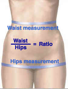
Yesterday, I wrote about a study that looked at a CT scan-derived index that promised to predict complications and mortality based on the waist-hip ratio. It was actually a very good one. But there is another abstract being presented at the American College of Surgeons Clinical Congress this week that promises miracles from the CT scanner as well.

This next abstract looks at muscle mass in trauma patients, as measured by CT scan. Specifically, the authors measured the density of the psoas muscle by determining its cross-sectional area and its density in Hounsfeld units. They then looked at the relationship between this and 90 day mortality, complications, and disposition location.

Really? Well, here are the factoids:

  • The study involved only 152 patients age 45+ from the year 2008
  • Median ISS was only 9
  • Patients with the lowest psoas cross-sectional area had an associated significantly higher death rate
  • Those with lowest psoas density had an associated increase in complications, dependency on discharge, and mortality
  • The authors suggest that these measurements could aid in patients who would benefit from aggressive nutritional support and physical therapy, and could aid in discharge planning

Bottom line: Very different from yesterday’s abstract. This one has no grounding in prior research. It appears to be one that was just dreamed up from nowhere. And it is truly an association study. No causality can or should be inferred.

There were only 152 patients studied. From 2008. Why? Why didn’t the authors use a more contemporary dataset? There is something weird going on behind the scenes. Is this an old study that was forgotten, and is just now being conveniently dusted off for analysis and submission? A power analysis to find out how many patients should be reviewed is not possible, so it is important to err on the high side. Not just 152 patients.

If you were to just read the abstract and especially the conclusions, you really might get the wrong idea. This is a study that will not see it’s day in any journal. Read and learn from it. But don’t duplicate it!

Related post:

Reference: Computed tomography-measured psoas density predicts complications, discharge location, and mortality in trauma patients. ACS Scientific Forum, trauma abstracts, 2016.


Using The CT Scan As A Crystal Ball For Trauma?

Two abstracts that are being presented at the American College of Surgeons Clinical Congress this week use CT scans to predict interesting things. They are things that you would not think a CT scanner should be able to do.

So can we use arcane measurements of things found on CT scan to make accurate predictions about our patients? Sure, if we see very low density bubbles (free air) in the abdomen, it’s pretty likely that some kind of abdominal catastrophe has occurred. Or if their is a large amount of high density fluid (blood) in the left chest after a stab wound, the patient will probably require a chest tube.

But what about other measurements that wouldn’t seem to be related to anything? Could we have found a magic crystal ball here, or is it just wishful thinking?

The first abstract evaluated the ability of the waist to hip ratio (WHR) to predict outcomes after trauma. Obviously, this is the width of the waist divided by the width of the body at the hips.

waist-hips-ratio1

Here are the factoids for this study:

  • 555 patients were analyzed retrospectively over 1 year at a Level I trauma center.
  • In-hospital complications and death were specifically analyzed
  • Using a receiver operating characteristic curve, the authors determined that a magic ratio of 1 was the best predictor
  • Complications were significantly higher in the group with WHR>1 (50% vs 19%) as was mortality (17% vs 7%)
  • Regression analysis showed that patients with WHR>1 were 4x more likely to have a complication and 3x more likely to die
  • WHR was only weakly correlated with BMI

Bottom line: WTF? How can this be, you ask? Just because your patient is a bit “fusiform” in shape, they have a rougher time after trauma? Well, in this case there may actually be some basis for the findings. There are thousands of articles in the literature that have identified that this shape actually is associated with higher complications and mortality in general. And there are already some published trauma papers that have confirmed this association. Interestingly, the BMI was less predictive that the WHR in this study, so this may be a better surrogate measure for obesity.

The number of patients enrolled is reasonable, and the statistics look sound (for just being an abstract). So there may be something here. However, before you start using the “measure tool” on your CT console on every trauma patient, wait for the confirmatory prospective studies to come along. 

Tomorrow, a look at a not-so-good study of this type, looking at an even more arcane metric on the CT scan.

Reference: Computed tomorgraphy-measured waist to hip ratio: a reliable predictor of outcomes after trauma. ACS Scientific Forum, trauma abstracts, 2016.

ED Use of CT – Everyone Does It Differently

There is tremendous variability in ordering imaging in trauma patients. To some degree, this is due to the dearth of standards pertaining to radiographic imaging, at least in trauma. And when standards do exist, trauma professionals are not very good at adhering to them. We’d rather do it our way. Or the way we were trained to do it.

The group at Jamaica Hospital in Queens, NY quantified some of those differences, studying ordering patterns of trauma surgeons (TS), emergency physicians (EP), and surgery chief residents (CR). Unfortunately, they then tried to draw some interesting conclusions, which I’ll discuss at the end.

They reviewed all blunt trauma activations over a 6 month period at their urban trauma center. At the end of each trauma activation, each of the three physician groups wrote imaging orders, but only the trauma surgeons’ were submitted. Missed injuries were defined as any that would not have been found based on each provider group’s orders. Extremity injuries, and those found on physical exam or plain imaging were excluded.

Here are the factoids:

  • The authors do not state how many patients they saw in this period, but by extrapolation it appears to be about 250
  • Trauma surgeons ordered significantly more studies (1,012) than the EPs (882) or CRs (884)
  • This resulted in essentially a “pan-scan” in 78%, 64%, and 69%, respectively
  • Radiation exposure was said to be the same for all groups (18 vs 13 vs 15 mSv) [I’m having a hard time buying this]
  • But cost was higher in the trauma surgeon group ($344 vs $267 vs $292) [Huh? Is this only the electric bill for the CT scanner? Very low, IMHO]
  • And the trauma surgeons had a missed injury rate of only 1%, vs 11% for EPs and 7% for CRs [Wow!]

Bottom line: Sorry, I just can’t believe these results. There are a lot of things left unsaid in this poster. What were all these missed injuries? What magical CT scan that only the trauma surgeons ordered actually picked them up? And probably most importantly, were they clinically significant? A small hematoma somewhere doesn’t make a difference (see the “tree falls in a forest” post below).

It looks to me like the authors wanted to justify their use of pan-scan, and push their emergency physicians to follow suit. Unfortunately, this is a poster presentation, meaning that there will be limited opportunity to question the authors about the specifics.

The debate regarding pan-scan vs selective imaging is an active one. The evidence is definitely not in yet. While we sort it out, the best path is to develop a reasonable imaging practice guideline based on the literature, where available. Some areas such as head and cervical spine CT have been worked out fairly well. Then fill in the blanks and encourage all trauma professionals in your hospital to follow them. There is great value in adhering to good guidelines, even when there are blanks in our knowledge.

Related posts:

Reference: Variability in computed tomography imaging of trauma patients among emergency department physicians and trauma surgeons with respect to missed injuries, radiation exposure and cost. AAST 2016, Poster #75.

IV Contrast and Trauma

We use CT scanning in trauma care so much that we tend to take it (and its safety) for granted. I’ve written quite a bit about thoughtful use of radiographic studies to achieve a reasonable patient exposure to xrays. But another thing to think about is the use of IV contrast.

IV contrast is a hyperosmolar solution that contains some substance (usually an iodine compound) that is radiopaque to some degree. It has been shown to have a significant impact on short-term kidney function and in some cases can cause renal failure.

Here are some facts you need to know:

  • Contrast nephrotoxicity is defined as a 25% increase in serum creatinine, usually within the first 3 days after administration
  • There is usually normal urine output and minimal to no proteinuria
  • In most cases, renal function returns to normal after 3-4 days
  • Nephrotoxicity almost never occurs in people with normal baseline kidney function
  • Large or repeated doses given within 72 hours greatly increase risk for toxicity
  • Old age and pre-existing diabetic renal impairment also greatly increase risk

If you must give contrast to a patient who is at risk, make sure they are volume expanded (tough in trauma patients), or consider giving acetylcysteine or using isosmolar contrast (controversial, may still cause toxicity).

Bottom line: If you are considering contrast CT, try to get a history to see if the patient is at risk for nephrotoxicity. Also consider all of the studies that will be needed and try to consolidate your contrast dosing. For example, you can get CT chest/abdomen/pelvis and CT angio of the neck with one contrast bolus. Consider low dose contrast injection if the patient needs formal angiographic studies in the IR suite. Always think about the global needs of your patient and plan accordingly (and safely).

Reference: Contrast media and the kidney. British J Radiol 76:513-518, 2003.

More On CT Usage In Unstable Patients

Yes, it is practically dogma that CT should not be used in unstable trauma patients. Either they go directly to the OR, or an attempt to stabilize them is briefly undertaken in the trauma bay. And as you know, I’m not a big believer in dogma. But this one has withstood the test of time. You can see my comments about a previous paper below in the related posts.

But now some authors in Colombia have published a paper that seems to call this idea into question. Could it be true? Read carefully!

This was a small, retrospective review of patients from a large Level I government designated trauma center. They reviewed their experience over a two year period, identifying all hemodynamically unstable patients in the registry. They excluded dead patients, those with isolated head injury, and any who had surgery at an outside hospital prior to transfer.

Here are the factoids:

  • 171 patients were reviewed, and of course they tended to be young males
  • 91 went straight to the OR, and 80 were taken to CT first
  • “Unstable” patients were defined as having SBP < 100 and/or HR > 100
  • Mechanism of injury for the OR group was 95% penetrating, but for the CT group was about 50:50 penetrating/blunt
  • The mean SBP and HR for the “unstable” patients taken to CT were 92 and 110, respectively
  • Mortality was the same for both groups (18% OR vs `13% CT)

Bottom line: The authors concluded that it is permissible to take unstable patients to CT if you don’t spend too much time there based on similar mortality rates. But the problem was that I don’t consider their patients to have been unstable! Mean SBP in their “unstable” group was over 90 torr and the heart rate was only 110! The lowest SBP was only 79. And mortality is too crude of an outcome to rely on. Furthermore, the patients they took to CT tended to have blunt mechanisms, and may not have had ample efforts at resuscitation in the trauma bay first, or may have met criteria to go to CT anyway (see related posts below).

Reference: Computed tomography in hemodynamically unstable severely injured blunt and penetrating trauma patients. J Trauma 80(4):597-603, 2016.