Tag Archives: ct scan

Routine CT After Operative Exploration For Penetrating Trauma

CT scans are commonly used to aid the workup of patients with blunt trauma. They are occasionally useful in penetrating trauma, specifically when penetration into a body cavity is uncertain and the patient has no hard signs that would send him or her immediately to the operating room.

Is there any role in operative penetrating trauma, after the patient has already been to the OR? The dogma has always been that the eyeballs of the surgeon in the OR are better than any other imaging modality. Really?

The surgical group at San Francisco General addressed this question by retrospectively reviewing 6 years of their operative penetrating injury registry data. They were interested in finding how many occult injuries (seen with CT but not by the surgeon) were found on a postop CT. A total of 225 patients who underwent operative management of penetrating abdomen or chest injury were included.

Here are the factoids:

  • Only 110 patients had a postop CT scan; 73 had scans within the first 24 hours, the other 37 were scanned later
  • Rationale for early scan was to investigate retroperitoneal injury in half of patients, but frequently no indication was given (41%)
  • Rationale for late scan was for workup of ileus in one third, or for evaluation of new or unexpected clinical problems
  • Occult injuries were found in about half of early CT patients (52%), and 22% of late CT patients
  • The most common occult injuries were fractures, GU issues, regraded solid organ injury, and unrecognized vascular injuries
  • Several management changes occurred, including

Bottom line: There appears to be a significant benefit to sending some penetrating injury patients to CT in the early postop period. Specifically, those with injury to the retroperitoneum, deep into the liver, near the spine, or with multiple and complicated injuries would benefit. Simple stabs and gunshots that stay away from these areas/structures probably do not need followup imaging. 

Rreference: Routine computed tomography after recent operative exploration for penetrating trauma: What injuries do we miss? J Trauma, published ahead of print, May 11, 2017.

How To: Manage Extraperitoneal Bladder Rupture

Extraperitoneal bladder rupture is a relatively uncommon injury, but is easily managed in most cases. It is associated with a blunt mechanism, and concomitant fracture of the pubic rami or spreading of the symphysis pubis is nearly always present. In the old days, we used to think that the bladder injury was due to penetration anteriorly by bony fragments, but this is probably an old wives tale. It’s more likely due to hydraulic forces occurring within the bladder at the same time the pelvic ring is being deformed or spread apart by blunt forces.

If you obtain a pelvic x-ray during the initial trauma evaluation and see any fractures or diastasis around the symphysis, think bladder injury. Placement of a urinary catheter will typically drain plenty of urine, which will usually be grossly bloody.

Once the injury is suspected, the diagnostic test of choice is a CT cystogram. Don’t confuse this with the images seen when the bladder passively fills with contrast when the catheter is clamped. There is not enough pressure in the bladder to guarantee that contrast will leak out, so this type of study may be falsely negative.

True CT cystogram technique requires filling the bladder with at least 350cc of dilute contrast under pressure by hanging it on an IV pole, then clamping the catheter. Once the bladder is filled, the scan can proceed as usual. But after it is complete, a second limited scan through the pelvis must be performed after the contrast has been evacuated by unclamping the catheter. This allows visualization of small contrast leaks that might otherwise be masked by all the contrast in the bladder.

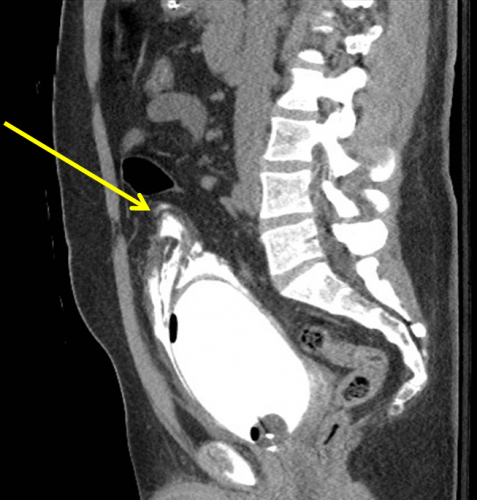
Here’s a nice sagittal image of an extraperitoneal injury from radiologypics.com:

Note how the contrast dissects around the bladder but does not enter the peritoneal cavity.

Extraperitoneal injuries usually do not require repair and will heal on their own. However, if the symphysis pubis needs instrumentation to restore anatomic position, concomitant repair of the bladder is frequently necessary to keep the hardware from being contaminated by urine.

Bottom line:

  • Suspect an extraperitoneal bladder injury in anyone with bony injuries involving the symphysis pubis.
  • Don’t order a urinalysis in trauma patients!
  • Use CT cystogram technique to make the diagnosis.
  • Treatment is simple: leave the urinary catheter in place for 10 days. No urology consult is needed.
  • Then repeat the CT cystogram to confirm healing, and remove the catheter.

Related posts:

EAST 2017 #2: CT Scan After Recent Operative Exploration for Penetrating Trauma

The general rule for penetrating trauma, especially gunshots to the abdomen, is that you don’t need to obtain a CT scan to help you decide to go to the OR. (Of course, there are a few exceptions.) And the corollary has always been that you don’t need to get a CT scan after you operate for penetrating trauma.

But the group at UCSF is questioning this. They retrospectively looked at 5 years of data on patients who underwent trauma laparotomy without preoperative imaging. They focused on new findings on CT that were not reported during the initial operation.

Here are the factoids:

  • 230 of 328 patients undergoing a trauma lap did not have preop imaging
  • 85 of the 230 patients (37%) underwent immediate postop CT scan. These patients tended to have a gunshot mechanism and higher injury severity score.
  • Unreported injuries were found in 45% (!) and tended to be GU and orthopedic in nature
  • 47% of those with unreported injuries found required some sort of intervention

Bottom line: This is a very interesting and potentially practice changing study. However, there is some opportunity for bias since only select patients underwent postop scanning. Nevertheless, one in five patients who did get a postop scan had an injury that required some sort of intervention. This study begs to be reworked to further support it, and to develop specific criteria for postop scanning.

Questions/comments for the authors/presenters:

  • Be sure to break down your results by gunshot vs stab. This will help formulate those criteria I mentioned above.
  • Specifically list the occult injuries and interventions required. In some studies, those “required interventions” are pretty weak (urology consult vs an actual procedure).
  • How exactly did the operating surgeons determine who to send to CT? Was it surgeon-specific (i.e. one surgeon always did, another never did)? Was it due to operative findings (hole near the kidney)? This is also needed when developing specific criteria for postop imaging.
  • Nice poster!

Click here to go the the EAST 2017 page to see comments on other abstracts.

Related posts:

Reference: Routine tomography after recent operative exploration for penetrating trauma: what injuries do we miss?  Poster #14, EAST 2017.

EAST 2017 #1: Accuracy of CT Scans Done Outside The Trauma Center

Imaging prior to transfer to a trauma center has been the subject of debate for years. The focus has primarily been on the necessity of these scans, and the sheer numbers that are done. For the most part, the discussion has been driven by the potential for radiation exposure.

This paper, from the University of Oklahoma, takes a different approach. The authors looked at the accuracy and adequacy of imaging performed prior to transfer to their Level I trauma center.

Patients were enrolled prospectively over an 8 month period in 2012. Outside images were interpreted by a single radiologist who was blinded to the outside interpretation.  If images were repeated, they were compared to the first scan, and the reason for the redo was noted.

Here are the factoids:

  • 235 consecutive transfer patients were enrolled, and 203 who had at least one CT scan were included in the final dataset
  • 76% of these patients had additional imaging performed once they arrived at the trauma center
  • Reasons for additional images were insufficient workup (76%) and technical inadequacy (31%)
  • Missed injuries were detected on outside CT scans 49% of the time, and the majority of them (90%) were deemed clinically significant
  • Missed injuries on a repeated scan were present in 54% of patients, and 76% were clinically significant
  • Overall, 73% of images (either outside or repeat) contained additional injuries

Bottom line: This is a new approach to assessing the value of outside imaging prior to transfer to a trauma center. I have always recommended that trauma centers work with their referral partners to assure them we don’t need them to do the workup for us. I encourage them to obtain only what they need to decide if they can keep the patient. But once they find anything that they cannot treat, stop all workup and prepare to transfer.

Questions/comments for the authors/presenters:

  • Why did you use such an old dataset?
  • Is this a prospective enrollment/retrospective analysis study designed to use an existing, old dataset?
  • How did you decide that outside imaging represented an inadequate workup? Do you have a diagnostic imaging guideline that you follow?
  • What are the credentials for your trauma radiologist?
  • How did you determine that a missed injury was clinically significant? Be sure to provide a list and explanation during your presentation.
  • Be sure to separate out missed injuries seen on the original CT from new missed injuries seen on the repeat scan.
  • Congratulations on looking at an old problem in a new way!

Click here to go the the EAST 2017 page to see comments on other abstracts.

Related posts:

Reference: Adequacy and accuracy of non-tertiary trauma center computed tomography: what are we missing? Paper #7, EAST 2017.

Pan Scanning for Elderly Falls?

The last abstract for the Clinical Congress of the American College of Surgeons that I will review deals with doing a so-called “pan-scan” for ground level falls. Apparently, patients at this center have been pan-scanned for years, and they wanted to determine if it was appropriate.

This was a retrospective trauma registry review of 9 years worth of ground level falls. Patients were divided into young (18-54 years) and old (55+ years) groups. They were included in the study if they received a pan-scan.

Here are the factoids:

  • Hospital admission rates (95%) and ICU admission rates (48%) were the same for young and old
  • ISS was a little higher in the older group (9 vs 12)
  • Here are the incidence and type of injuries detected:
Young (n=328) Old (n=257)
TBI 35% 40%
C-spine 2% 2%
Blunt Cereb-vasc inj * 20% 31%
Pneumothorax 14% 15%
Abdominal injury 4% 2%
Mortality * 3% 11%

 * = statistically significant

Bottom line: There is an ongoing argument, still, regarding pan-scan vs selective scanning. The pan-scanners argue that the increased risk (much of which is delayed or intangible) is worth the extra information. This study shows that the authors did not find much difference in injury diagnosis in young vs elderly patients, with the exception of blunt cerebrovascular injury.

Most elderly patients who fall sustain injuries to the head, spine (all of it), extremities and hips. The torso is largely spared, with the exception of ribs. In my opinion, chest CT is only for identification of aortic injury, which just can’t happen from falling over. Or even down stairs. And solid organ injury is also rare in this group.

Although the future risk from radiation in an elderly patient is probably low, the risk from the IV contrast needed to see the aorta or solid organs is significant in this group. And keep in mind the dangers of screening for a low probability diagnosis. You may find something that prompts invasive and potentially more dangerous investigations of something that may never have caused a problem!

I recommend selective scanning of the head and cervical spine (if not clinically clearable), and selective conventional imaging of any other suspicious areas. If additional detail of the thoracic and/or lumbar spine are needed, specific spine CT imaging should be used without contrast.

Related posts:

Reference: Pan-scanning for ground level falls in the elderly: really? ACS Surgical Forum, trauma abstracts, 2016.