Tag Archives: ct scan

IV Contrast and Trauma – Revisited

We use CT scanning in trauma care so much that we tend to take it (and its safety) for granted. I’ve written quite a bit about thoughtful use of radiographic studies to achieve a reasonable patient exposure to xrays. But another thing to think about is the use of IV contrast.

IV contrast is a hyperosmolar solution that contains some substance (usually an iodine compound) that is radiopaque to some degree. It has been shown to have a significant impact on short-term kidney function and in some cases can cause renal failure.

Here are some facts you need to know:

  • Contrast nephrotoxicity is defined as a 25% increase in serum creatinine, usually within the first 3 days after administration
  • There is usually normal urine output and minimal to no proteinuria
  • In most cases, renal function returns to normal after 3-4 days
  • Nephrotoxicity almost never occurs in people with normal baseline kidney function
  • Large or repeated doses given within 72 hours greatly increase risk for toxicity
  • Old age and pre-existing diabetic renal impairment also greatly increase risk

If you must give contrast to a patient who is at risk, make sure they are volume expanded (tough in trauma patients), or consider giving acetylcysteine or using isosmolar contrast (controversial, may still cause toxicity).

Bottom line: If you are considering contrast CT, try to get a history to see if the patient is at risk for nephrotoxicity. Also consider all of the studies that will be needed and try to consolidate your contrast dosing. For example, you can get CT chest/abdomen/pelvis and CT angio of the neck with one contrast bolus. Consider low dose contrast injection if the patient needs formal angiographic studies in the IR suite. And finally, consider what changes will be made if the study is positive. For example, if a CT angio of the neck for blunt carotid/vertebral injury is being considered, the intervention for a positive result is usually just aspirin. Since this is a very benign medication, why not forgo the scan and just start aspirin if there is a significant risk of kidney injury from the contrast. Always think about the global needs of your patient and plan accordingly (and safely).

Reference: Contrast media and the kidney. British J Radiol 76:513-518, 2003.

Radiation Exposure From Imaging At Adult vs Pediatric Trauma Centers

Anyone who reads this blog already knows I am a big believer in well-crafted and focused practice guidelines. And by focused I mean directed toward a clinical problem that typically sees a lot of variability between care providers. Use of imaging is one of these clinical problems. A surgeon may order a certain set of studies for a major blunt trauma patient, and their emergency medicine colleague might order a somewhat different set for someone with the exact same history, physical exam, and injury pattern. Who is right? Neither!

And the variability is even greater when we throw a pediatric patient into the mix. Trauma professionals tend to be even more “generous” when ordering studies on children because they are afraid they might miss something. Unfortunately, this has the potential for overuse of imaging and exposure to unnecessary radiation.

Avery Nathens and a consortium of pediatric trauma centers used the Trauma Quality Improvement Database (TQIP) to review CT imaging practices on children age < 18 over a four year period. Only blunt trauma patients were studied, and the Abbreviated Injury Scale had to be at least 2 for a minimum of one organ system. Transfer patients were excluded because there is no data on imaging for the referring hospital in the TQIP database for them. Comparisons were made between practices at adult trauma centers treating children (ATC), mixed adult/pediatric centers (MTC) and pediatric only trauma centers (PCT).

Here are the factoids:

  • Over 59,000 pediatric trauma patients were identified in the data, and about half (31,081) received at least one CT scan
  • The distribution among the three types of trauma centers was even, with roughly a third seen at each
  • Of the study group 46% had a head CT, 17% a chest CT, and 26% underwent abdominal CT
  • Injured children were more likely to undergo CT if they were older, had a higher ISS, lower motor GCS, were involved in a car crash, or had severe injuries to head or torso
  • Overall CT rates were about the same across the three types of centers (56% ATC, 57% MTC, 43% PTC)
  • Chest CT was performed 8x as much at ATC/MTC vs PTC (!)
  • Abdominal CT was performed 2x as much at ATC/MTC vs PTC
  • Lesser injured children received relatively more CT scans at ATC/MTC when compared to PTC
  • Using standard estimates of cancer risk from all CT scans received, children treated at adult or mixed trauma centers received enough radiation to cause 17 additional lifetime cancers per 100,000 patients
  • About 35 additional lifetime cancers per 100,000 would be caused by the chest and abdominal scans performed at the ATC/MTC centers when compared to pediatric-only centers

Bottom line: This is yet another reason to adopt a well-designed pediatric imaging guideline. Not only are adult centers using CT scanning much more that pediatric-only centers, but they are unnecessarily adding to the lifetime risk for cancer of our children!

As I always recommend, find a well-designed imaging guideline from an established pediatric center and “borrow” it. Sure, it may need a few minor tweaks to fit well with your hospital. That’s okay. Just get it done so your team can begin to order the initial imaging studies consistently and intelligently.

Reference: Computed tomography rates and estimated radiation-associated cancer risk among injured children treated at different trauma center types. Injury 2018, in press.  https://doi.org/10.1016/j.injury.2018.09.036

Pan Scanning for Elderly Falls?

The last abstract for the Clinical Congress of the American College of Surgeons that I will review deals with doing a so-called “pan-scan” for ground level falls. Apparently, patients at this center have been pan-scanned for years, and they wanted to determine if it was appropriate.

This was a retrospective trauma registry review of 9 years worth of ground level falls. Patients were divided into young (18-54 years) and old (55+ years) groups. They were included in the study if they received a pan-scan.

Here are the factoids:

  • Hospital admission rates (95%) and ICU admission rates (48%) were the same for young and old
  • ISS was a little higher in the older group (9 vs 12)
  • Here are the incidence and type of injuries detected:
Young (n=328) Old (n=257)
TBI 35% 40%
C-spine 2% 2%
Blunt Cereb-vasc inj * 20% 31%
Pneumothorax 14% 15%
Abdominal injury 4% 2%
Mortality * 3% 11%

 * = statistically significant

Bottom line: There is an ongoing argument, still, regarding pan-scan vs selective scanning. The pan-scanners argue that the increased risk (much of which is delayed or intangible) is worth the extra information. This study shows that the authors did not find much difference in injury diagnosis in young vs elderly patients, with the exception of blunt cerebrovascular injury.

Most elderly patients who fall sustain injuries to the head, spine (all of it), extremities and hips. The torso is largely spared, with the exception of ribs. In my opinion, chest CT is only for identification of aortic injury, which just can’t happen from falling over. Or even down stairs. And solid organ injury is also rare in this group.

Although the future risk from radiation in an elderly patient is probably low, the risk from the IV contrast needed to see the aorta or solid organs is significant in this group. And keep in mind the dangers of screening for a low probability diagnosis. You may find something that prompts invasive and potentially more dangerous investigations of something that may never have caused a problem!

I recommend selective scanning of the head and cervical spine (if not clinically clearable), and selective conventional imaging of any other suspicious areas. If additional detail of the thoracic and/or lumbar spine are needed, specific spine CT imaging should be used without contrast.

Related posts:

Reference: Pan-scanning for ground level falls in the elderly: really? ACS Surgical Forum, trauma abstracts, 2016.

Part 2: Metal Splints – Can You CT Scan Through Them?

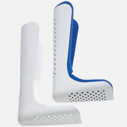
In my last post, I debunked the myth that using a pre-formed aluminum splint significantly degrades the quality of standard x-rays. But what about a study that provides much more detail, such as CT scan?

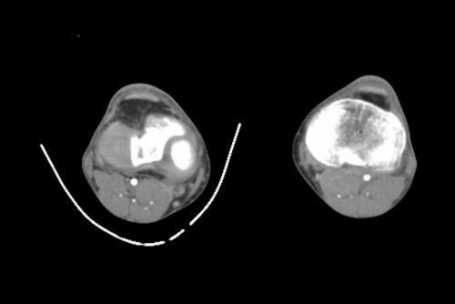
CT scan techs have told me that there would be too much artifact using any kind of metal splint. And typically, when imaging an extremity with CT, we are looking at vascular runoff. The vessels are small, and high image quality is extremely important. If the images are bad, then we risk having to give the patient another dose of both radiation and contrast.

As you know, my mantra is question everything! So i scouted around and found some images to share using one of these splints. Look closely for the intimal flap in the image below:

Can’t see it? That’s because it isn’t there! But you certainly could if it were!

Bottom line: A perforated aluminum splint causes absolutely no artifact or image degradation. Do not cause additional injury by removing it prior to imaging, either CT or conventional x-ray. Although your friendly techs, radiologists, and orthopedic surgeons may moan, it won’t hurt their ability to make decisions on the images.

ACS Trauma Abstract #6: Scanning Unstable Trauma Patients???

If you’ve read my stuff for very long, you know I frown on sending unstable patients anywhere but to the OR. Instability tends to get worse, and that always happens at inopportune locations like hallways, elevators, and CT scanners. Imagine my surprise when I noticed an abstract being presented at the Clinical Congress of the American College of Surgeons this week suggesting that it was okay to scan hemodynamically unstable patients before “definitive therapy.”

Here’s the title:

“Computed tomography in hemodynamically abnormal thoracoabdominal trauma safely enhances surgical triage”

The devil is in the details and the language. This group from USC included all patients who were hemodynamically abnormal on arrival to the trauma bay but who normalized to SBP > 90 during the resuscitation were included.  A total of 253 of these patients were reviewed over a 9 year period, and the usual variables were analyzed (mortality, complications, hospital, ICU, and vent days, etc).

Here are the factoids:

  • Of the 253 patients studied, 45 went to straight to OR and 208 were taken to CT
  • Injury severity was identical for the two groups
  • Lengths of stay and mortality were not different, but only p values were given
  • Patients taken to CT cleared their lactic acidosis faster (12 vs 5 hours), and used a bit less plasma and significantly less blood transfusions
  • The OR group underwent more procedures (31% vs 13%), although what these were and when they were performed is not listed

Bottom line: The title of this abstract is misleading, and may fool those who don’t read the rest of the abstract. It should read:

“Computed tomography in previously hemodynamically abnormal thoracoabdominal trauma safely enhances surgical triage”

Someone who just skims through this issue of the journal may get the idea that it’s okay to scan an unstable patient. The authors are not saying this at all. If you read the conclusion carefully, you can see that the patients had to be resuscitated to a SBP > 90 before they considered taking to scan. And they did that for the majority of these patients.

The real question is, why do the scanned patients clear their lactic acidosis faster, need less blood, and undergo fewer procedures? It appears that there is some bias or selection process in play. Otherwise, why not use the magic CT scanner to make them all better?

Reference: Computed tomography in hemodynamically abnormal thoracoabdominal trauma safely enhances surgical triage. JACS 225(4S2):e175-176, 2017.