Tag Archives: complications

AAST 2019 #5: DOACs Part 1

A short while ago I wrote about the proper nomenclature of the new or novel oral anticoagulant medications that are replacing warfarin in patients with atrial fibrillation (click here for details). Cut to the chase, the consensus seems to be that they should be called direct oral anticoagulants or DOACs.

These medications strike fear into the average trauma professional, primarily because there is no easy way to reverse them as there is for warfarin. We are finally accumulating enough experience with them to start to see the bigger picture with respect to complications and mortality. Today, I’ll begin the discussion with a series of three abstracts regarding these drugs.

The AAST conducted a multicenter, prospective, observational study that collected DOAC trauma patient information from 15 centers. They reviewed four years of data, specifically examining the use of reversal agents and mortality.

Here are the factoids:

  • A total of 606 patients were enrolled. They were generally elderly with an average age of 75.
  • Most were taking one of the Factor Xa inhibitors (apixaban, rivaroxaban, edoxiban), while just 8% were taking the direct thrombin inhibitor dabigatran.
  • Only 1% of patients received a reversal agent (prothrombin complex concentrate (PCC) 87%, Praxbind (12%), and Andexxa (1%)
  • Those receiving reversal tended to be older than the average and had more severe head injuries
  • Patients who were reversed with PCC had no change in mortality using a regression model
  • Patients reversed with Praxbind or Andexxa had a 15x higher probability of mortality

The author’s conclusions merely restated their results.

This is fascinating information. Unfortunately, this study was not designed to provide a comparison with patients taking warfarin. However, my next two abstract reviews will cover this very topic. 

There are two interesting tidbits here. First, reversal was only carried out in about one in eight patients. Why is this? No protocol? No product? Too pricey? Patients not hurt badly enough? And how would that be judged anyway?

The second is that reversal with PCC seems to be benign, but use of one of the specifically designed reversal agents really jacked up mortality. These agents (Praxbind and Andexxa) are very expensive ($3.5K and $50K respectively). Furthermore, there are no studies anywhere that show their effectiveness. This one actually seems to show they might be dangerous.

The devil is in the details. Here are my questions for the presenter and authors:

  • Were there any guidelines for reversal? This is key because if not, the statistics just describe “how we do it.” Yes, you can tease out higher ISS or AIS head as potential reasons, but were there directions regarding this built into the study protocol?
  • Do you have any data on the success rates of PCC reversal? Were there provisions to demonstrate lesion stability vs progression after administration?
  • Do you have an impression of why the tailored reversal agents seemed to be so deadly? Were they used as a last resort due to cost. Did the centers have a hard time getting it or authorizing its use?

This abstract could be a gold mine!

Reference: The AAST prospective, observational, multicenter study investigating the initial experience with reversal ofnovel oral anticoagulants in trauma patients. AAST 2019, Oral Paper 58.

Trocar Chest Tubes Or Blunt Technique? Part 2

In my last post on chest tube insertion technique, I reviewed a paper that compared chest tube insertion complications using two different trocar tips, blunt plastic and sharp metal. The sharp tip tubes caused more complications, although the study was weakened by the fact that the physicians inserting the tubes were complete newbies.

Today, I’ll discuss what the authors call a “best evidence topic” that reviewed the safety of the trocar technique. It is similar to a meta-analysis of available literature that attempts to reach a conclusion regarding this type of tube insertion. A literature search from 1946 to 2013 was conducted seeking to pull all papers on trocar chest tube insertion techniqes. A total of 258 papers were identified, but on closer inspection only 7 were identified that “provided the best evidence to answer the question.”

Here are the factoids from some of these papers:

  • Tube malposition occurred significantly more often in a series of 106 trocar tubes inserted into 75 ICU patients
  • In trocar tubes inserted for trauma, CT showed malplacement in 29% vs 19% with non-trocar tubes [This latter number seems very high to me!]
  • A retrospective study of 1249 patients resulted in the trocar technique being abandoned due to severe lung and stomach injuries
  • Use of trocar technique was associated with a significantly higher incidence of re-expansion pulmonary edema in 92 patients with spontaneous pneumothorax
  • A poorly controlled prospective study showed 23 complications with trocar technique and none with blunt dissection. The denominator could not be determined.

Bottom line: Overall, the literature is just not good enough to answer this question. But it does provide some suggestions.

  • Trocar insertion can be done well in experienced hands. Cardiac surgeons use these all the time, although sometimes they have the benefit of already being in the chest so they can visualize the point of entry and control the tip.
  • Any chest tube insertion can go awry.  It’s very important to learn proper technique, and take care to apply it faithfully, even in emergency situations.
  • If you really like trocars and want to improve insertion safety, start with the blunt dissection technique first, sweep a finger inside the chest to ensure there are no adhesions, then insert the trocar tube to guide it into position. Please note that I do not believe that we can control the tube once the instrument (trocar or clamp) are removed from the chest. And the tube will work fine just about anywhere it ends up (unless that’s the spleen).
  • Newbies should be supervised carefully and learn blunt insertion technique first. Be mindful that it is still possible to pass the insertion clamp into the same structures as a trocar if you are not careful. My practice is to place my fingers about 2 cm from the tip of the clamp as I push it through the pleura. If the pleura gives way more easily than anticipated, by fingers will keep the clamp from going too far into the chest. 
  • Always mark your insertion spot before prepping. This will generally be lateral to the nipple in men, so always prep the nipple into your field as a landmark.
  • Always be careful!

Reference: Is the trocar technique for tube thoracostomy safe in the current era? Interactive CV and thoracic surg 19:125-128, 2014.

Trocar Chest Tubes Or Blunt Technique? Part 1

This is an old question: what is the best way to insert a chest tube? There are several techniques available to us:

  • Blunt dissection and insertion
  • Trocar with a blunt tip (plastic stylet)
  • Trocar with a sharp tip (metal stylet)
  • Seldinger technique for small tubes

Typically, when there are multiple ways to do a thing, then there is no clear choice as to which is better. It then becomes a personal choice, or one driven by the financial considerations of the equipment used, and demonstrates the need for a practice guideline.

There are very few good papers out there that critically compare any of these techniques. Today, I’ll review one cadaver study and tomorrow I’ll tackle one “best evidence” paper that attempt to answer it.

A group in Vienna, Austria performed a cadaver study comparing the use of the two types of trocar tubes:

The top tube is the sharp trocar type, the bottom is the blunt trocar.

The study engaged twenty emergency medicine residents who had little, if any, experience placing chest tubes. Each placed 10 chest tubes (5 of each type) in fresh cadavers after undergoing a one-hour standardized lecture on anatomy, technique, and complications. The authors tabulated insertion times, as well as complication and success rate based on anatomic dissection.

Tube type was randomly assigned for each attempt by each resident. One blunt insertion and one sharp insertion were performed on opposite sides of a cadaver each month for the trainees. Over a period of 5 months, each resident performed 10 total insertions.

Here are the factoids:

  • Mean time to insertion for blunt vs sharp tips was the same, about 60 seconds
  • Insertion time declined by about 20 seconds by the final attempt at 5 months
  • Accurate placement occurred in 94% of blunt tip tubes vs 86% of sharp tip tubes
  • There were significantly more complications with the sharp tip (4 below diaphragm, 5 outside the thorax, 1 in the liver,  and 4 in the spleen) vs the blunt tip (2 below diaphragm, 2 extrathoracic, 2 in the liver, and 2 aborted due to damage to the tube)
  • BMI did not increase complications, but it did increase insertion time significantly

The authors concluded that there is a 6-14% complication rate that is operator related, and that the incidence of complications was increased with the use of a sharp tip tube. They warn against the use of these tubes.

Bottom line: This is certainly an interesting study. The insertion numbers are sort of reasonable, and the use of fresh cadavers is okay. They are not quite as realistic as real living people, but close. The biggest drawback was that they used chest tube newbies, most of whom had never inserted a tube. And they were placed in the unrealistic setting where they had to attend training and watch a video, then insert two tubes per month without coaching or supervision. This is not how we do it in the real world. 

I was impressed with what I consider the high number of complications. I don’t typically see that many, although I work at a blunt dissection institution. However, it does show that any trocar style tube is probably more like a weapon in inexperienced hands. So perhaps, even with supervision, both sharp and blunt trocar types should be avoided in the teaching setting. Sure, blunt dissection may take a bit longer, but the tube is also less likely to end up somewhere it shouldn’t be.

Tomorrow: Review of a “best evidence” review from New York.

Reference: Evaluation of performance of two different chest tubes with either a sharp or a blunt tip for thoracostomy in 100 human cadavers. Scand J Trauma Resus Emerg Med 20:10, 2012.

Air Embolism From an Intraosseous (IO) Line

IO lines are a godsend when we are faced with a patient who desperately needs access but has no veins. The tibia is generally easy to locate and the landmarks for insertion are straightforward. They are so easy to insert and use, we sometimes “set it and forget it”, in the words of infomercial guru Ron Popeil.

But complications are possible. The most common is an insertion “miss”, where the fluid then infuses into the knee joint or soft tissues of the leg. Problems can also arise when the tibia is fractured, leading to leakage into the soft tissues. Infection is extremely rare.

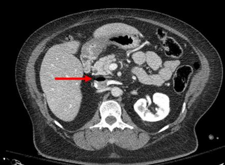
This photo shows the inferior vena cava of a patient with bilateral IO line insertions (black bubble at the top of the round IVC).

During transport, one line was inadvertently disconnected and probably entrained some air. There was no adverse clinical effect, but if the problem is not recognized and the line is not closed properly, there could be.

Bottom line: Treat an IO line as carefully as you would a regular IV. You can give anything through it that can be given via a regular IV: crystalloid, blood, drugs. And even air, so be careful!

Best of AAST #8: Complications After Trauma Laparotomy

With the introduction of damage control laparotomy (DCL) in the early 1990s, the trauma literature has focused on the nuances of this procedure. A significant amout of research has looked at patient selection, techniques, optimum time to closure, and complications afterwards. Studies on the single-look trauma laparotomy (STL) seem to have fallen behind. When compared to DCL, it seems to have relatively few complications.

But is that really so? A paper from the 1980s showed a nearly 50% complication rate after STL, but this included some trivial things like atelectasis which padded the numbers. A group at Scripps Mercy in San Diego looked at long-term complications after  STL in a state-wide California database. They were able to identify patients who underwent STL who were then readmitted for complications at a later date. They studied this data over an 8-year period.

Here are the factoids:

  • A total of 2,113 patients had a STL during the study period
  • One third (712) were readmitted at least once, with a median time to first readmission of 110 days
  • 30% of these patients had a surgery-related complication:
    • bowel obstruction 18%
    • infection 9%
    • incisional hernia 7%
  • Mechanism of injury was not related to development of complications

Bottom line: More than 10% of patients undergoing single-look trauma laparotomy develop significant complications. This is much higher than the complication rate seen after typical general surgical procedures. The difference between these groups and the reasons are not clear. Additional work must be done to tease out the risk factors, and our patients should be counseled on these potential complications and when to return for evaluation. Finally, the trauma surgeon should always use their best judgment to avoid an unnecessary trauma laparotomy.

Reference: Long-term outcomes after single-look trauma laparotomy: a large population-based study. Session IV Paper 14, AAST 2018.