Tag Archives: chest tube

Practice Guideline: Chest Tube Management (Part 2)

In my last post, I went over the rationale for developing a practice guideline for something as simple and lowly as chest tube management. Today, I’m posting the details of the guideline that’t been in use at my hospital for the past 15 years. I’ve updated it to reflect two lessons learned from actually using it.

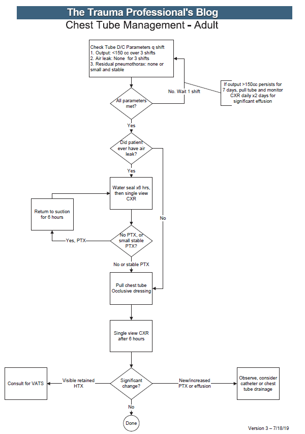
Here’s an image of the practice guideline. Click to open a full-size copy in a new window:
Here are some key points:

  • Note the decision tree format. This eliminates uncertainty so that the clinician can stick to the script. There are no hedge words like “consider” used. Just real verbs.
  • We found that hospital length of stay improved when we changed the three parameters from daily monitoring to three consecutive shifts. We are prepared to pull the tube on any shift, not just during the day time. And it also allows this part of the guideline to be nursing driven. They remind the surgeons that criteria are met so we can immediately remove the tube.
  • Water seal is only used if there was an air leak at some point. This allows us to detect a slow ongoing leak that may not be present during our brief inspection of the system on rounds.
  • The American College of Surgeons Committee on Trauma expects trauma centers to monitor compliance with at least some of their guidelines. This one makes it easy for a PI nurse or other personnel to do so.
  • The first of the “new” parts of this guideline is: putting a 7 day cap on failure due to tube output greater than 150cc per three shifts. At that point, the infectious risks of keeping a tube in begin to outweigh its efficacy. Typically, a small effusion may appear the day following removal, then resolves shortly.
  • The second “new” part is moving to VATS early if it is clear that there is visible hemothorax that is not being drained by the system. Some centers may want to try irrigation or lytics, but the data for this is not great. I’ll republish my posts on this over the next two days.

Click here to download a copy of this practice guideline for adults.

Click here to download the pediatric chest tube practice guideline.

Practice Guideline: Chest Tube Management (Part 1)

I’m devoting the next series of posts to revisiting the management of hemo- and/or pneumothorax. These clinical issues are some of the most common sources of variability in how trauma professionals approach them. Let’s start with the seemingly simple chore of managing a lowly chest tube.

Management of chest tubes is one of those clinical situations that are just perfect for practice guideline development: commonly encountered, with lots of variability between trauma professionals. There are lots of potential areas for variation:

  • How long should the tube stay in?
  • What criteria should be used to determine when to pull it?
  • Water seal or no?
  • When should followup x-rays be done?

Every one of these questions will have a very real impact on that patient’s length of stay and potential for complications.

We developed a chest tube clinical practice guideline (CPG) at Regions Hospital way back in 2004! Of course, there was little literature available to guide us in answering the questions listed above. So we had to use the clinical experience and judgment of the trauma faculty to settle on a protocol that all were comfortable with.

Ultimately, we answered the questions like this:

  • The tube stays in until three specific criteria are met
  • The criteria are: <150 cc drainage over 3 shifts, no air leak, and no residual pneumothorax (or at least a small, stable one)
  • Use of water seal is predicated on whether there was ever an air leak
  • An x-ray is obtained to determine whether any significant pneumo- or hemothorax is present prior to pulling the tube, and 6 hours after pulling it

This CPG has been in effect for over 15 years with excellent results and dramatically shortened lengths of stay.  However, as with any good practice guideline, it needs occasional updates to stay abreast of new research literature or clinical experiences. We recognized that occasional patients had excessive drainage for an extended period of time. This led us to limit the length of time the tube was in to seven days. And we also noted that a few patients had visible hemothorax on their pre-pull imaging. These patients were very likely to return with clinical symptoms of lung entrapment, so we added a decision point to consider VATS at the end of the protocol.

I’ll share the full protocol tomorrow and provide a downloadable copy that you can modify for your own center. I’ll also give a little more commentary on the rationale for the key decision points in this CPG.

Related posts:

Best Of AAST 2021: Chest Tube Based On Pneumothorax Size

How big is too big? That has been the question for a long time as it applies to pneumothorax and chest tubes. For many, it is a math problem that takes into account the appearance on chest x-ray, the physiology of the patient, and their ability to tolerate the pneumothorax based on any pre-existing medical conditions.

The group at Froedtert in Milwaukee has been trying to make this decision a bit more objective. They introduced the concept of CT based size measurement using a 35mm threshold at this very meeting three years ago. Read my review here. My criticisms at the time centered around the need to get a CT scan for diagnosis and their subjective definition of a failure requiring chest tube insertion. The abstract never did make it to publication.

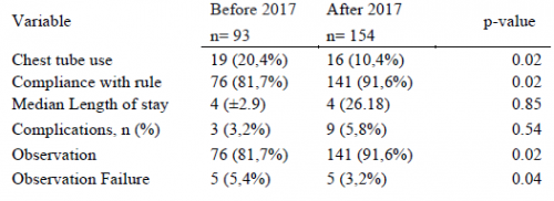
The authors are back now with a follow-on study. This time, they made a rule that any pneumothorax less than 35mm from the chest wall would be observed without tube placement. The performed a retrospective review of their experience and divided it into two time periods: 2015-2016, before the new rule, and 2018-2019, after the new rule. They excluded any chest tubes inserted before the scan was performed, those that included a sizable hemothorax, and patients placed on a ventilator or who died.

Here are the factoids:

  • There were 93 patients in the early period and 154 in the later period
  • Chest tube use significantly declined from 20% to 10% between the two periods
  • Compliance with the rule significantly increased from 82% to 92%
  • There was no difference in length of stay, complications, or death
  • Observation failure was marginally less in the later period, and statistical significance depends on what method you use to calculate it
  • Patients in the later group were 2x more likely to be observed (by regression analysis)

The authors concluded that the 35mm rule resulted in a two-fold increase in observation and decreased the number of unnecessary CT scans.

Bottom line: I still have a few issues with this series of abstracts. First, decision to insert a chest tube requires a CT scan in a patient with a pneumothorax. This seems like extra radiation for patients who may not otherwise fit any of the usual blunt imaging criteria. And, like their 2018 abstract, there is no objective criteria for failure requiring tube insertion. So the number of insertions can potentially be quite subjective based on the whims of the individual surgeon.

What this abstract really shows is that compliance with the new rule increased, and there were no obvious complications from its use. The other numbers (chest tube insertions, observation failure) are just too subjective to learn much from.

Here are my questions for the presenter and authors:

  • Why was there such a large increase in the number of subjects for two identical-length time periods? Both were two years long, yet there were two-thirds more patients in the later period. Did your trauma center volumes go up that much? If not, could this represent some sort of selection bias that might change your numbers?
  • You concluded that your new rule decreased the number of “unnecessary” CT scans? How so? It looks like you are using more of them!
  • Do you routinely get a chest CT on all your patients with pneumothorax? Seems like a lot of radiation just to decide whether or not to put a tube in.
  • How do you manage a pneumothorax found on chest x-ray? Must they get a CT? Or are you willing to watch them and follow with serial x-rays?
  • How do you decide to take out the chest tube? Hopefully not another scan!

There should be some very interesting discussion of this abstract!

Reference: THE 35-MM RULE TO GUIDE PNEUMOTHORAX MANAGEMENT: INCREASES APPROPRIATE OBSERVATION AND DECREASES UNNECESARY CHEST TUBES. AAST 2021, Oral abstract #56.

Chest Tube Repositioning – Final Answer

So you’re faced with a chest tube that “someone else” inserted, and the followup chest xray shows that the last drain hole is outside the chest. What to do?

Well, as I mentioned, there is very little written on this topic, just dogma. So here are some practical tips on avoiding or fixing this problem:

  • Don’t let it happen to you! When inserting the tube, make sure that it’s done right! I don’t recommend making large skin incisions just to inspect your work. Most tubes can be inserted through a 2cm incision, but you can’t see into the depths of the wound. There are two tricks:
    • In adults with a reasonable BMI, the last hole is in when the tube markings show 12cm (bigger people need bigger numbers, though)
    • After insertion, get into the habit of running a finger down the radiopaque stripe on the tube all the way to the chest wall. If you don’t feel a hole (which is punched through the stripe), this will confirm that the it is inside, and that the tube actually goes into the chest. You may laugh, but I’ve seen them placed under the scapula. This even looks normal on chest xray!
  • Patients with a high BMI may not need anything done. The soft tissue will probably keep the hole occluded. If there is no air leak, just watch it.
  • If the tube was just put in and the wound has just been prepped and dressed, and the hole is barely outside the rib line, you might consider repositioning it a centimeter or two. Infection is a real concern, so if in doubt, go to the next step.
  • Replace the tube, using a new site. Yes, it’s a nuisance and requires more anesthetic and possibly sedation, but it’s better than treating an empyema in a few days.

Chest Tube Repositioning – Part 2

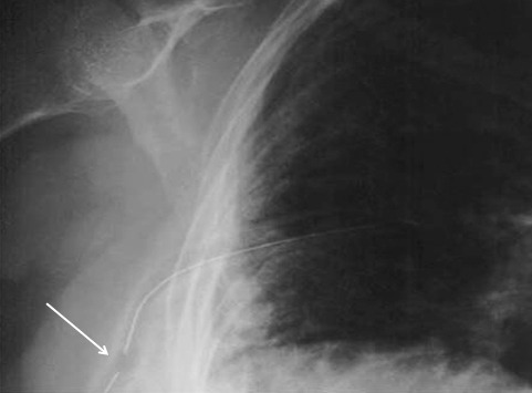
Yesterday I presented the problem of the malpositioned chest tube, specifically one that is not completely in the pleural space. This one is way out:

So what do the doctor books say? Well, the first thing you will discover if you try to look it up is that THERE IS NO LITERATURE ON THIS COMMON PROBLEM! There are a few papers on tubes placed in the fissure and tubes inserted into the lung parenchyma. But there are only a few mentions of tubes with holes still outside the chest.

I’ve gotten a number of comments, including “you can push them in a little”, “take it out and put in another”, and “never push them in.” Since we don’t have any science to guide us, we have to use common sense. But remember, I’ve shown you plenty of examples where something seems reasonable, but turns out to be ineffective or downright harmful.

There are three principles that guide me when I face this problem:

  • Prevention is preferable to intervention
  • Do no (or as little as possible) further harm
  • Be creative

Tomorrow, I’ll finish this series and provide some tips and guidelines to help manage this problem using the principles outlined above.