Tag Archives: AI

New Technology: Using AI To Interpret Pelvic X-rays

Look out, radiologists! The computers are coming for you!

Radiologists use their extensive understanding of human anatomy and combine it with subtle findings they see on x-ray shadow pictures. In doing this, they can identify a wide variety of diseases, anomalies, and injuries. But as we have seen with vision systems and game playing (think chess), computers are getting pretty good at doing this as well.

Is it only a matter of time until computer artificial intelligence (AI) starts reading x-rays?  Look at how good they already are at interpreting EKGs. The trauma group at Stanford paired up with the Chang Gung Memorial Hospital in Taiwan to test the use of AI for interpreting images to identify a specific set of common pelvic fractures.

The Stanford group used a deep learning neural network (XCeption) to analyze source x-rays (standard A-P pelvis images) from Chang Gung. These x-rays were divided into training and testing cohorts. The authors also applied different degrees of blurring, brightness, rotation, and contrast adjustment to the training set in order to help the AI overcome these issues when interpreting novel images.

The AI interpreted the test images with a very high degree of sensitivity, specificity, accuracy, and predictive values, with all of them over 0.90. The algorithms generated a “heat map” that showed the areas that were suspicious for fracture. Here are some examples with the original x-ray on the left and the heat map on the right:
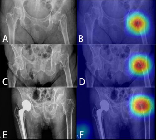
The top row shows a femoral neck fracture, the middle row an intertrochanteric fracture, and the bottom row another femoral neck fracture with a contralateral implant. All were handily identified by the AI.

AI applications are usually only as good as their training sets. In general, the bigger the better so they can gain a broader experience for more accurate interpretation. So it is possible that uncommon, subtle fractures could be missed. But remember, artificial intelligence is meant to supplement the radiologist, not replace him or her. You can all breathe more easily now.

This technology has the potential for broader use in radiographic interpretation. In my mind, the best way to use it is to first let the radiologist read the images as they usually do. Once they have done this, then turn on the heat map so they can see any additional anomalies the AI has found. They can then use this information to supplement the initial interpretation.

Expect to see more work like this in the future. I predict that, ultimately, the picture archiving and communications systems (PACS) software providers will build this into their product. As the digital images are moving from the imaging hardware to the digital storage media, the AI can intercept it and begin the augmented interpretation process. The radiologist will then be able to turn on the heat map as soon as the images arrive on their workstation.

Stay tuned! I’m sure there is more like this to come!

Reference: Practical computer vision application to detect hip fractures on pelvic X-rays: a bi-institutional study.  Trauma Surgery and Acute Care Open 6(1), http://dx.doi.org/10.1136/tsaco-2021-000705.

Best Of EAST 2023 #8: Use Of AI To Detect Rib Fractures On CT

Artificial intelligence systems (AI) are increasingly finding their way into medical practice. They have been used to assist pathologists in screening microscope specimens for years. Although still amazingly complicated, one of the most obvious applications for trauma is in reading x-rays. Counting rib fractures may be helpful for care planning, and characterizing fracture patterns may assist our orthopedic colleagues in evaluating and planning rib plating procedures.

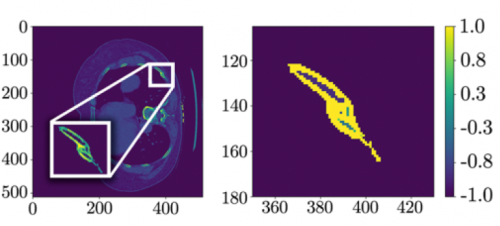
The trauma group at Stanford developed a computer vision system to assist in identifying fractures and their percent displacement.  They used a variation on a neural network deep learning system and trained it on a publicly available CT scan dataset.  They used an index of radiographic similarity (DICE score) to test how well their model matched up against the reading of an actual radiologist.

Here are the factoids:

  • The AI network was trained on a dataset of 5,000 images in 660 chest CT scans that had been annotated by radiologists
  • The model achieved a DICE score of 0.88 after training
  • With a little jiggering of the model (reweighting), the receiver operating characteristic curve improved to 0.99, which is nearly perfect

The left side shows a CT scan rotated 90 degrees; the right side shows the processed data after a fracture was detected.

Bottom line: This paper describes what lies ahead for healthcare in general. The increasing sophistication and accuracy of AI applications will assist trauma professionals in doing their jobs better. But rest easy, they will not take our jobs anytime soon. What we do (for the most part) takes very complex processing and decision making. It will be quite some time before these systems can do anything more that augment what we do.

Expect to see these AI products integrated with PACS viewing systems at some point in the not so distant future. The radiologist will interpret images in conjunction with the AI, which will highlight suspicious areas on the images as an assist. The radiologist can then make sure they have reported on all regions that both they and the AI have flagged.

Here are my questions and comments for the presenter/authors:

  • How can you be sure that your model isn’t only good for analyzing your training and test datasets? If neural networks are overtrained, they get very good at the original datasets but are not so good analyzing novel datasets. Have you tried the on your own data yet?
  • Explain what “class reweighting” is and how it improved your model. I presume you used this technique to compensate for the potential issue mentioned above. But be sure to explain this in simple terms to the audience.
  • Don’t lose the audience with the net details. You will need to give a basic description of how deep learning nets are developed and how they work, but not get too fancy.

This is an interesting glimpse into what is coming to a theater near you, so to speak. Expect to see applications appearing in the next few years.

Reference: AUTOMATED RIB FRACTURE DETECTION AND CHARACTERIZATION ON COMPUTED TOMOGRAPHY SCANS USING COMPUTER VISION. EAST 2023 Podium paper #16.

Best Of EAST #17: Artificial Intelligence vs TRISS

The TRISS score is the great grand-daddy of probability of survival prediction in trauma, first introduced in 1981. It is a somewhat complicated equation that takes the injury severity score (ISS), revised trauma score (RTS), and age and cranks out a probability between 0 and 100%. Over the years, this system has been well validated, and its shortcomings have been elucidated as well.

Many authors have attempted to develop a system that is better than TRISS. Years ago, there was the New-TRISS. And back in the day (early 1990s) I even developed a neural network to replace TRISS. In general, all of these systems may improve accuracy by a few percent. But it has never been enough to prompt us to ditch the original system.

The group at the University of California at Los Angeles developed a machine learning algorithm using ICD-10 anatomic codes and a number of physiologic variables to try to improve upon the original TRISS score. They analyzed three years of NTDB data and attempted to predict in-hospital survival.

Here are the factoids:

  • The authors used over 1.4 million records to develop their model
  • Overall, 97% of patients survived, and survivors tended to be younger, have higher blood pressure, and have sustained a blunt mechanism (no surprises here at all)
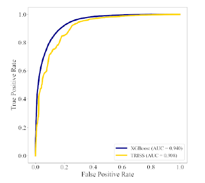
  • The ROC C-statistic for the false positive rate was better with the machine learning model (0.940 vs 0.908), as was the calibration statistic (0.997 vs 0.814)

Here is the ROC curve for machine (blue) vs TRISS (yellow):

The authors conclude that the machine learning model performs better than TRISS and that it may improve stratification of injury.

Bottom line: This study is one of many attempting to improve upon good old TRISS probability of survival. Why have there been so many attempts, and none that have appeared to “stick?” Here are my thoughts:

  • They are complicated. Sure, the original TRISS equation is slightly complicated, but it’s nothing close to a machine AI algorithm.
  • The inner workings are opaque. It’s not very easy to “open the box” and see which variables are actually driving the survival calculations.
  • The results are only as good as the training data. There is a real skew toward survival here (97%), so the algorithm will more likely be right in guessing that the patients will survive.
  • The improvements in these systems are generally incremental. In this case the ROC value increases from .908 to .940. Both of these values are very good.

In general, any time a new and better algorithm is introduced that shows much promise, someone wants to patent it so they can monetize the work.  Obviously, I don’t know anything about the plans for this algorithm. Somehow I doubt that many centers would be willing to abandon TRISS for an incremental improvement that may not be clinically significant at any price.

Here are my questions for the authors and presenter:

  • Please detail how you selected the variables to enter into the machine learning algorithm. Were they chosen by biased humans who had some idea they might be important, or did the AI comb the data and try to find the best correlations?
  • Be sure to explain the ROC and calibration statistics well. Most of the audience will be unfamiliar.
  • Are you using your model in your own performance improvement program now? If so, how is it helping you? If not, why?

Fascinating paper! Let’s here more about it!

Reference: ICD-10-BASED MACHINE LEARNING MODELS OUTPERFORM THE TRAUMA AND INJURY SEVERITY SCORE (TRISS) IN SURVIVAL PREDICTION, EAST 35th ASA, oral abstract #38.

New Technology: Using AI To Interpret Pelvic X-rays

Look out, radiologists! The computers are coming for you!

Radiologists use their extensive understanding of human anatomy and combine it with subtle findings they see on x-ray shadow pictures. In doing this, they can identify a wide variety of diseases, anomalies, and injuries. But as we have seen with vision systems and game playing (think chess), computers are getting pretty good at doing this as well.

Is it only a matter of time until computer artificial intelligence (AI) starts reading x-rays?  Look at how good they already are at interpreting EKGs. The trauma group at Stanford paired up with the Chang Gung Memorial Hospital in Taiwan to test the use of AI for interpreting images to identify a specific set of common pelvic fractures.

The Stanford group used a deep learning neural network (XCeption) to analyze source x-rays (standard A-P pelvis images) from Chang Gung. These x-rays were divided into training and testing cohorts. The authors also applied different degrees of blurring, brightness, rotation, and contrast adjustment to the training set in order to help the AI overcome these issues when interpreting novel images.

The AI interpreted the test images with a very high degree of sensitivity, specificity, accuracy, and predictive values, with all of them over 0.90. The algorithms generated a “heat map” that showed the areas that were suspicious for fracture. Here are some examples with the original x-ray on the left and the heat map on the right:

The top row shows a femoral neck fracture, the middle row an intertrochanteric fracture, and the bottom row another femoral neck fracture with a contralateral implant. All were handily identified by the AI.

AI applications are usually only as good as their training sets. In general, the bigger the better so they can gain a broader experience for more accurate interpretation. So it is possible that uncommon, subtle fractures could be missed. But remember, artificial intelligence is meant to supplement the radiologist, not replace him or her. You can all breathe more easily now.

This technology has the potential for broader use in radiographic interpretation. In my mind, the best way to use it is to first let the radiologist read the images as they usually do. Once they have done this, then turn on the heat map so they can see any additional anomalies the AI has found. They can then use this information to supplement the initial interpretation.

Expect to see more work like this in the future. I predict that, ultimately, the picture archiving and communications systems (PACS) software providers will build this into their product. As the digital images are moving from the imaging hardware to the digital storage media, the AI can intercept it and begin the augmented interpretation process. The radiologist will then be able to turn on the heat map as soon as the images arrive on their workstation.

Stay tuned! I’m sure there is more like this to come!

Reference: Practical computer vision application to detect hip fractures on pelvic X-rays: a bi-institutional study.  Trauma Surgery and Acute Care Open 6(1), http://dx.doi.org/10.1136/tsaco-2021-000705.