Tag Archives: abstract

Best Of AAST 2021: Liposomal Bupivacaine For Rib Fractures

The mainstays of rib fracture management are pain control and pulmonary toilet. The pain part of the equation can be managed in many ways, using topical, oral, IV, and injectable medications.

Rib blocks have been a mainstay for achieving some degree of local pain control. Classically, xylocaine was injected in the area around the costal nerve at or proximal to the fracture site. Then we found that if we combined the anesthetic agent with epinephrine, we could prolong the effect. New, longer-acting agents came around, and we could achieve a longer duration of action.

Then there is the new kid on the block: liposomal bupivacaine, also known as Exparel in the US.  The manufacturer was able to take molecules of bupivacaine and encapsulate them in a lipid membrane. When injected, these little liposomes slowly release their cargo, with a more prolonged anesthetic effect. Allegedly.

Sounds great! But does it work? The group at University of Cincinnati designed a prospective, double-blinded, randomized placebo control study of liposomal bupivacaine vs saline injection for pain control in up to six rib fractures. Subjects had significant injury as measured by their inability to achieve at least 50% of the desired inspiratory capacity. The authors monitored a number of respiratory parameters, as well as the pain score.

Here are the factoids:

  • Two cohorts of 50 patients were recruited, one received liposomal bupivacaine in up to six rib fractures, and the other received saline injections
  • The bupivacaine group achieved higher incentive spirometry volumes over the first two days, by about 200 cc
  • There was no change in daily pain scores in either group
  • Both groups showed a similar decrease in opioid use over time
  • Hospital and ICU lengths of stay were the same, and there were no complications or adverse events

Bottom line: Hmm. What’s going on here? There is a moderate amount of literature out there that does indicate a positive effect from liposomal bupivacaine in other conditions. But there are also some blinded, randomized studies that fail as well. So there are three possibilities:

  1. Liposomal bupivacaine isn’t a panacea, and works better in some situations than others
  2. This study failed to show a real difference for some reason
  3. A combination of both

This is a relatively small study, and the authors were not able to share their power analysis. They did not state if the spirometry volumes were significantly different, although I’m not sure 200 cc is clinically relevant. Maybe. But pain scores remained similar and opioid use declined as expected in both. 

These kinds of studies can be important. The difference in cost between injecting liposomal bupivacaine ($19 / ml) vs regular bupivacaine (10 cents / ml) vs saline/nothing (free) is striking. The premium price for the liposomal form needs to have a clear benefit or a cheaper product should be used.

Here are my questions for the presenter and authors:

  • Was your study big enough to show a result? Show us your power analysis.
  • How significant was the incentive spirometry result. Was the difference clinically noticeable?
  • What is your takeaway for this study? Your conclusion parrots the results. What will you do differently now, if anything?

Reference: INTERCOSTAL LIPOSOMAL BUPIVACAINE INJECTION FOR
RIB FRACTURES. AAST 2021, Oral abstract #20.

Best Of AAST 2021: Identifying Risk For Elderly Falls

Over the past 20 years, falls have become the most common mechanism of injury at most trauma centers. In fact, many centers count twice as many falls as motor vehicle crashes! The problem with working in a trauma center is that we tend to see patients at risk for falls only after they have fallen.

The group at Butterworth Hospital attempted to determine if there was a way to identify patients at risk for falls earlier. They postulated that many of these patients may have experienced a fall within the past year, identifying them as at high risk for yet another. They retrospectively reviewed their trauma registry data for a three year period. Specifically, they wanted to identify how many of those had suffered earlier falls and what happened to them over time.

Here are the factoids:

  • A total of 597 patients were also admitted due to a fall during the year prior to their index admission
  • Only 2% had falls prevention teaching after the previous admission
  • About a third of patients fell again within a year after the index admission, and 20% were admitted again
  • The patients were assessed using the Hester-Davis score (see below), and patients who were identified by it as high risk were more likely to be readmitted or die
  • Overall mortality at 12 months was about 20%

The authors were surprised that so many of their falls patients had been previously admitted for a fall. They recognized that it presents a major prevention opportunity, and recommend these patients undergo some type of activity before and/or after discharge.

Note: The Hester Davis Fall Risk Scale (HDFRS) includes factors of age, date of last known fall, mobility, medications, mental status, toileting needs, volume/electrolyte status, communication/sensory, and behavior with the option to choose multiple options per risk category; a score of seven to ten indicates low fall risk, eleven to fourteen indicates moderate fall risk, and greater than fifteen indicates high fall risk.

Bottom line: This is a straightforward single-hospital registry study. Even though it reflects the experience of a single rural trauma center, the results are applicable to most others. It confirms that any fall in the elderly should be considered a sentinel event which has a good chance leading to death within a year. 

Here’s the way I see it:

“You fall, you die”

It is very important that every trauma center identify these patients when they arrive, and apply prevention efforts while in the hospital or hook them up with activities after discharge. And if you don’t have such a program included in your injury prevention activities, you should! It’s the most common mechanism seen by trauma centers, hands down!

I have only one suggestion for the presenter and authors:

  • The concept of being “at risk” was not clear to me. Did this mean that you looked back one year for each admission to see if there was an admission for a fall? Or did you just get the history of a fall from the previous admission? It looks like you identified an index admission, then looked back a year to see if the patient should be included in this study. Then you looked forward a year to see if there was yet another admission and/or death. Is this correct? Please clarify during your presentation at the meeting.

Reference:  FALL RISK IDENTIFICATION THROUGHOUT THE
CONTINUUM OF CARE. AAST 2021, Oral abstract #18.

Best Of AAST 2021: Reducing Errors In Trauma Care

Finally, a performance improvement (PI) abstract at AAST!

As many of you know, there are two general types of issues that are encountered in the usual PI processes: provider (peer) vs system. Provider issues are errors of omission or commission by an individual clinician. Examples include a surgeon making a technical error during a procedure, or prescribing the wrong drug or dose for some condition.

One might think that provider issues are the most common type of problem encountered. But they would be wrong. The vast majority of clinicians go to work each day with the idea that they will do their job to the best of their abilities. So how could things go awry?

Because the majority of errors have some degree of system component! They are set up to fail by factors outside their perception and/or control. Let’s look at a surgeon who has several small bowel anastomoses fall apart. His surgery department head chides/educates him, reports him to hospital quality, and proctors his next ten bowel cases. Everything is good, right?

But then, two months later, the stapler company issues a recall because they found a higher than usual number of anastomotic failures with one of their products. So it wasn’t the surgeon after all, like everyone assumed. This is an extreme example, but you get the idea. System issues often look like peer issues, but it’s frequently difficult for many PI programs to recognize or accept this.

A multi-institutional group reviewed the results of a newly implemented Mortality Reporting System (MRS) to analyze a large number of PI opportunities for improvement (OFI). More than 300 trauma centers submitted data to the MRS when a death occurred where an OFI was identified. The reports included details of the incident and mitigation strategies that were applied.


Here are the factoids:

  • A total of 395 deaths were reviewed over a two year period
  • One third of deaths were unanticipated (!!), and a third of those were failure to rescue
  • Half of errors pertained to clinical management, clinical performance, and communication
  • Human failures occurred in about two thirds of cases
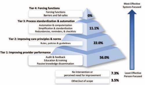
  • The most common remedy applied was education, which presumes a “provider issue”
  • System strategies like automation, standardization, and fail-safe approaches were seldom used, implying that system issues were seldom recognized
  • in 7%, the trauma centers could not identify a specific strategy to prevent future harm (!!!)

The authors concluded that most strategies to reduce errors focus on individual performance and do not recognize the value of system-level intervention.

Bottom line: Look at the pyramid chart above (interesting choice for a chart, but very effective). The arrow shows progression from provider focus to systems focus. The pyramid shows how the recognition of and intervention for system issues drops off very rapidly.

I am both shocked and fascinated by the last bullet point. A strategy couldn’t be developed to prevent the same thing from happening again. Now, there are a few rare instances where this could be correct. Your patient could have been struck by a bolt of lightning in her room, or a meteorite could have crashed through the wall. But I doubt it. This 7% illustrates the importance of investigating all the angles to try to determine how the system failed!

For once, I have no critique for an abstract. It is a straightforward descriptive study that reveals an issue that many in PI are not fully aware of. I’ll definitely be listening to this one, and I really look forward to the published paper!

Reference: ERROR REDUCTION IN TRAUMA CARE: LESSONS FROM AN ANONYMIZED, NATIONAL, MULTI-CENTER MORTALITY REPORTING SYSTEM. AAST 2021, Oral abstract #17.

Best Of AAST 2021: Hard Signs Of Vascular injury

Well, it’s that time of year again! The annual American Association for the Surgery (AAST) is just a few weeks away. Starting today, I will begin reviewing some of the interesting abstracts (to me, at least) that will be presented. I’ll give my analysis and perspective, and usually provide some questions for the presenters that they may face during the live meeting. Enjoy!

I’ll start with abstract #1. This one is from the AAST Prospective Observational Vascular Injury Trial (PROOVIT).  The group was established to create an aggregate database of information on the presentation, diagnosis, management (acute and definitive), surveillance and outcomes following vascular trauma. It manages a registry that collects a wide variety of data on assorted vascular injuries.

This study re-examines our use of “hard signs” to diagnose vascular injury. Back in the day, we had “hard signs” and “soft signs.”  Hard signs were fairly obvious indicators of serious injury, such as pulselessness, ischemia, pulsatile bleeding, expanding hematoma, or a thrill or bruit. Soft signs were a bit less harsh: history of arterial bleeding, diminished pulse, stable hematoma, or an injury in proximity to the vessel.

In the old days, any hard sign of vascular injury was a hard indication to proceed directly to the OR for exploration and repair. However, the authors argue that in this day and age of advanced imaging and noninvasive treatment, maybe hard signs aren’t as hard as they used to be. They postulated that distinguishing between hemorrhage and ischemia would be more important in determining management of these injuries.

They focused on femoral and popliteal artery injuries, searching the database for classic hard vs soft signs, and newer ischemic (absent or diminished pulses, frank limb ischemia) vs hemorrhagic signs (overt hemorrhage, expanding hematoma, hypotension). They examined the presentation, pathology, treatment and outcome in 521 patients in the registry

Here are the factoids:

  • Hard signs occurred in 386, and 35% underwent CT angio instead of moving directly to OR
  • Soft signs occurred in the remaining 175, and 39% went to the OR without any further imaging
  • When using hemorrhage (HEM) vs ischemia (ISC), there were significant differences in mechanism (more penetrating in HEM), incidence of concomitant vein and nerve injury (higher in HEM), transection (higher in HEM), occlusion (higher in ISC)
  • For diagnosis and management, HEM was more likely to get intervention sooner, without imaging, using ligation or primary repair
  • ISC was more likely to undergo endovascular repair
  • HEM patients used a little more blood and had a higher mortality rate
  • Amputation rates, lengths of stay, and graft outcomes were the same

The authors concluded that the old hard vs soft signs paradigm no longer works, and suggest that using hemorrhage vs ischemia in now more useful.

Bottom line: This is a simple, straightforward descriptive study of five years of vascular injury of the proximal lower extremity. It certainly paints the picture that the old paradigm doesn’t work as well as it used to. About a third of patients with hard signs had preop imaging, and about the same number with soft signs went straight to OR.

The major drawback is that this is what I call a “how we do it study.” The results are largely dependent on the predominant practices at the participating centers. What if most of the centers that chose to participate are much more likely to use diagnostic imaging first, or go straight to OR first? And that centers that obeyed the classic hard vs soft signs paradigm steered clear? That could skew the results in this study.

This is a very thought-provoking paper. I’m looking forward to hearing more of the details at the meeting. I’ll be in the front row!

Questions for the authors and presenter:

  • Why did you focus only on femoral and popliteal injuries? What should be do about the others?
  • What were the “demographics” of the participating centers? What trauma center level, academic or not, urban or rural? All of these could have a significant impact on your numbers.
  • What was the duration of experience captured in the database? Are you able to see changes in preop eval or straight to OR practice over the years?

Reference: HARD, SOFT, & IRRELEVANT: HEMORRHAGIC & ISCHEMIC SIGNS BETTER DISTINGUISH IMPORTANT CHARACTERISTICS OF EXTREMITY VASCULAR INJURIES. AAST 2021, Oral abstract #1.


Best of AAST #10: Pediatric Contrast Extravasation And Pseudoaneurysms

There is a significant amount of variation in the management of pediatric solid organ injury. This is well documented between adult and pediatric trauma centers in t, but also apparently between centers in different countries. A poster from a Japanese group in Okinawa Japan will be presented this week detailing the relationship between contrast extravasation after spleen or liver injury and pseudoaneurysm formation.

In adults, the general rule is that pseudoaneurysms just about anywhere slowly enlarge and eventually rupture. This group sought to define this relationship in the pediatric age group. They performed a multi-center observational study of retrospectively enrolled children, defined as age 16 and less. Those who had contrast extravasation on initial CT were monitored for later pseudoaneurysm formation.

Here are the factoids:

  • 236 patients were enrolled across 10 participating centers, with about two-thirds having liver injury and the remainder with splenic injury
  • 80% of patients underwent followup CT scan (!!)
  • 33 patients (15%) underwent angiography (!!!!)
  • 17 patients with CT scan (2%) had pseudoaneurysm formation and 4 of them had a delayed rupture
  • Overall, pseudoaneurysms occurred in 29% of those with contrast extravasation and 5% without extravasation
  • The authors concluded that contrast extravasation was significantly associated with pseudoaneurysm formation after adjusting for variables such as ISS, injury grade, and degree of hemoperitoneum

Bottom line: This is an abstract, so a lot is missing. What was the age distribution, especially among those who underwent angiography? Was the data skewed by a predominantly teenage population, whose organs behave more like adults? The abstract answers a question but ignores the clinical significance.

For those trauma professionals who routinely care for pediatric patients, you know that contrast extravasation in children doesn’t act like its adult counterpart. Kids seldom decompensate, and for those who are mistakenly taken for angiography, the extravasation is frequently gone. The authors even admitted in the conclusion that aggressive screening and treatment for pseudoaneurysm was carried out.

The real question is, what is the significance of a solid organ pseudoaneurysm in children? Based on my clinical experience and reading of the US literature, not much. Of course, there is a gray zone as children move into adulthood in the early to mid-teens. But this does not warrant re-scanning and there should be no routine angiography in this age group. Contrast extravasation in pediatric patients warrants close observation for a period of time. But intervention should only be considered in those who behave clinically like they have ongoing bleeding. 

Reference: Association between contrast extravasation on CT scan and pseudoaneurysm in pediatric blunt splenic and hepatic injury: a multi-institutional observational study. Poster 31, AAST 2018.