Category Archives: What the heck?

What The Heck? CT Imaging Problem

Here’s one for you. A patient is brought to you after a motor vehicle crash. You’ve completed your evaluation in the trauma resuscitation room, and you move off to CT for some imaging.

As the techs are preparing to do the abdominal CT, they perform the scout image to set up the study. This is what you see:

The arm was left down due to a fracture (note the splint along the forearm). But what is all that debris on the image? Other than a few abrasions here and there, nothing is visible on the skin in those areas.

What the heck? What do you think these are? Will they interfere with imaging? And what can you do about it?

Tweet or comment with your answers. I will explain all tomorrow.

Print Friendly, PDF & Email

Consultant Gives An Unusual Recommendation: What Would You Do?

I know this has happened to most of you at one point or another:

One of your trauma patients sustains an injury outside of your area of expertise. You engage a consultant to evaluate that condition and manage it. They do so, and it requires some type of invasive procedure. They return from the procedure, and as you are rounding on the patient, you find the consultant has ordered a medication that you have not seen ordered for that procedure before.

What would you do? You are now in an interesting place. Do you discontinue the order? Call up the consultant and ask, what the heck? Might you poison your relationship with them in the process? And what is the impact on your patient?

Lots of questions, but here is what I recommend:

  • Hit the lit! Always assume that they might know something you don’t. They are an expert in their field for a reason, so give them the benefit of the doubt. Thoroughly review the literature to see if this is an approved new practice. But remember, a single interesting paper should never be enough to change your (or their) practice. There needs to be a sufficient body of literature showing that the practice is sound.
  • Talk to the consultant. Now that you are armed with the current thinking, ask them what they were thinking! Let them explain their rationale. Since you have already looked at the available data, you will be able to ask appropriate questions and deflect answers like, “well that’s how we did it where I trained.”
  • Change the orders. Assuming the order was not sound, it’s time to undo the ones that started this entire debate. Get rid of them now so you’re not stepping on any toes. However, if you believed that the order/medication would have been potentially harmful, don’t wait. You should have done it even before the first step!
  • Disseminate the info. Make sure that all of your partners are aware of the issue and the correct course of action (or orders). And send a note to the consultant group summarizing the discussion so none of your consultant’s partners make the same mistake again.

Tomorrow, a set of guidelines to give all of your consultants to make sure they behave appropriately and interface will with the trauma service.


Print Friendly, PDF & Email

What The Heck? Pigtail Catheter Chest Tube – The Answer

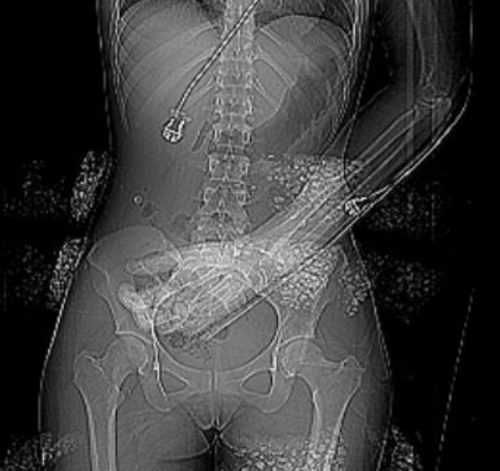
I previously described a trauma patient who had a pigtail type chest tube inserted with some odd CT findings after insertion:

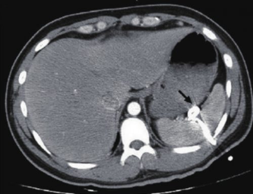
So what is wrong in this picture? Well, the catheter has been inserted into the spleen! This can occur if it is inserted too low, or if there are adhesions between lung and chest wall or diaphragm.

How can it be avoided? Make sure that the insertion point is no lower than the 5th intercostal space. This is the level of the nipple in a male. And depending on what type of kit you use, be careful! Some are based on Seldinger technique, which would seem to be a bit safer. Others use a small trochar, which can be inserted a little too deeply at times. Note that this complication can occur with any kit, and can also occur when using a standard tube and open insertion technique.

Does a pigtail tube even work for hemothorax? There’s some debate about this. Traumatic hemothorax is not defibrinated like a medical one. Thus, there are frequently clots present which may not fully evacuate through a standard chest tube, let alone a tiny one. Thus, I don’t recommend a pigtail for acute traumatic hemothorax.

How should I manage this issue? Obviously, this tube needs to come out. And assuming that the initial indication for the tube is still present, a better one needs to be inserted. Dont’ pull it out yet! First, look at the vital signs. If there is significant bleeding and/or vitals are not normal, an immediate trip to the operating room is in order. In this case, the patient will likely lose their spleen.

If vital signs are stable, book both an interventional radiology suite and an OR. Or better yet, use a hybrid room. Have the radiologist obtain a baseline angiogram, and position a catheter in the main splenic artery. Incrementally remove the pigtail, hand injecting a small amount of contrast each time. If extravasation is noted at any time, the radiologist can then attempt to embolize. If selective embolization isn’t successful, then the main splenic artery should be embolized. If embolization doesn’t work, or vital signs deteriorate at any time, the surgeon should immediately proceed to laparotomy. Attempts at splenic salvage will probably not be successful.

Finally, insert a new, conventional chest tube using finger guidance. Don’t make the same mistake twice! And by the way, this works for pigtails in the liver, too. They are less likely to bleed significantly when withdrawn, and obviously the radiologist can only used selective embolization if they do.

Print Friendly, PDF & Email

What The Heck? Pigtail Catheter Chest Tube

Here’s a case to make you think!

A patient arrives after being t-boned in his driver side door. He complains of left sided chest and abdominal pain. Chest x-ray shows a modest left hemopneumothorax. The decision is made to insert a pigtail type chest tube, and this is carried out in your trauma bay. It is uneventful, and a small amount of blood but no air is returned. The pelvis x-ray is unremarkable

The patient is then taken to CT, where an abdomen/pelvis scan with contrast is performed. This interesting slice is noted. What the heck?!

Here are my questions:

  • What is wrong in this picture?
  • How could it have been avoided?
  • Does a pigtail chest tube work for hemothorax?
  • How should this issue be managed, and where?

I’ll address these questions in my next post, and more!

Image source: internet

Print Friendly, PDF & Email

What The Heck? CT Imaging Problem: The Answer

I received some good guesses about this image yesterday, but no one got the right answer.

The patient had sustained blunt trauma and was undergoing CT imaging. The scout for the abdominal CT showed some kind of weird debris that interfered with the image, but when we uncovered and looked at the patient, nothing was visible:

What the heck? If you look carefully at the left side of the image, you can see that the “debris field” is on the surface of the patient. We can’t see in 3-D on images, but the difference in appearance on the left and right sides looks like it this stuff is wrapping around the patient.

She was brought in by EMS with a warming blanket in place. On closer inspection, this was a thin, disposable blanket that heats up when removed from an airtight plastic pouch. These blankets contain thin pockets of a mineral mixture that looks like gravel. When exposed to air it heats up.

But on CT it looks like bone density material! When we looked at the patient, we were just lifting off the blanket that contained the offending material. Hence, we couldn’t find it.

Here’s a picture of one of these products. Note the six mineral pouches embedded in it., Don’t let this happen to you!


Print Friendly, PDF & Email