Category Archives: What the heck?

What The Heck? CT Imaging Problem

Here’s one for you. A patient is brought to you after a motor vehicle crash. You’ve completed your evaluation in the trauma resuscitation room, and you move off to CT for some imaging.

As the techs are preparing to do the abdominal CT, they perform the scout image to set up the study. This is what you see:

The arm was left down due to a fracture (note the splint along the forearm). But what is all that debris on the image? Other than a few abrasions here and there, nothing is visible on the skin in those areas.

What the heck? What do you think these are? Will they interfere with imaging? And what can you do about it?

Tweet or comment with your answers. I will explain all tomorrow.

Print Friendly, PDF & Email

What The Heck? Final Answer: Progressive Back Pain After Heavy Lifting

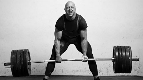
In my last two posts, I described an athlete who developed significant pain in his lower back after rapidly escalating his weight lifting regimen. The pain was very localized to the paraspinal areas bilaterally, and serum CPK was elevated.

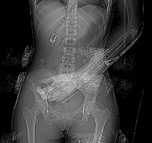
Congrats to Jay Slutsky for being the first to figure this one out. The suspected diagnosis was lumbar paraspinous muscle compartment syndrome. Compartment pressures were measured, and were found to be 78 and 26 mm Hg. A contrast MRI was obtained that showed swelling of both sets of paraspinal muscles.

The patient was taken to the OR for fasciotomy.
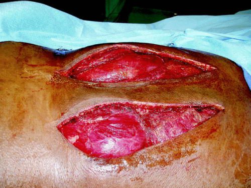
Source: Published paper

Note the bulging musculature above. Some areas appeared to be necrotic and did not bleed or contract. There were sharply debrided. The patient recovered quickly, with significant pain relief. The skin incisions were closed after several days, once swelling had subsided. He was well-healed and pain-free at his one month postop visit.

As you can see, any muscle surrounded by a rigid fascial compartment can develop a compartment syndrome. Typically, this requires direct trauma, but exertional compartment syndromes as in this case have been described in the legs of athletes as well. A history of a blow to the muscle group, or of very intense exercise should raise suspicion.

Physical findings of extreme pain that is very focal, coupled with discrete tenderness and firm muscle compartments, should confirm the potential diagnosis. Serum CPK is helpful for trending. Normal pressures in this muscle group tend to be in the single digits to low teens. They rise transiently during exercise, but usually return to normal shortly afterwards. “Normal” compartment pressures are not really known, so findings need to be coupled with CPK levels. Once the compartment pressure reaches the 30s, and especially if accompanied by high and rising CPKs, the syndrome is present. MRI is interesting, but not terribly helpful.

Treatment is typical for any compartment syndrome: release the muscle! A vertical incision centered over the bulging and tight muscle compartment is used. The wound is left open until swelling subsides enough to close the skin. Recovery is usually rapid, although some complain of a persistent low level of pain for a period of time. It is not known how soon these patients may resume sports or training.

Bottom line: Any patient with direct trauma or extreme exertion involving a muscle group is at risk for compartment syndrome. Physical exam, coupled with compartment pressure measurement if in doubt, are the mainstays of diagnosis. CPK levels may help in cases of uncertainty. As with any compartment syndrome, rapid diagnosis and fasciotomy is the key to preserving function and decreasing the likelihood of disability and chronic pain.

Related posts:

References:

  • Acute Exertional Lumbar Paraspinal Compartment Syndrome. Spine 35(25):E1529-E1533, 2010.
  • Lumbar paraspinal compartment syndrome. International Orthopaedics 36:1221-1227, 2011.
  • Paravertebral compartment syndrome after training causing severe back pain in an amateur rugby player: report of a rare case and review of the literature. BMC Musculoskelet Disord 14:259, 2013.


Print Friendly, PDF & Email

What The Heck? Part 2: Progressive Back Pain After Heavy Lifting

Yesterday, I described a case of a young athlete who developed progressive back pain after rapidly increased his deadlift weights. He presented to the hospital with back pain and inability to get up from a supine position. He had firm and tender paraspinal muscles in his lower back, but no other findings.

What to do next? Obviously, we need a bit more information on the bony structures. Other than run of the mill muscle strain, a compression fracture would be the next most common diagnosis. In this young, healthy athlete, a simple set of AP and lateral spine images should be sufficient. But if you opted for a CT scan, I won’t argue. In either case, the images were normal.

Since there is significant muscle pain and tenderness, a lab panel with a few extras is in order, as well. The usual electrolytes, etc were normal. Creatinine was 0.9, but CPK was 60,000!

Now what are you thinking? What’s the diagnosis, and what is the decision tree for treatment?

Add your comments below, or tweet them out. I’ll finish this topic up in the next post.


Print Friendly, PDF & Email

What The Heck? Progressive Back Pain After Heavy Lifting

What the heck?! Here’s an interesting case of back pain! Can you figure it out?

A 20 year old male athlete has been performing 125 pound deadlift exercises recently. During his last session, he rapidly escalated to 6 reps at 235 pounds. He developed crampy lower back pain two hours later. The pain became rapidly worse, and he was evaluated at a hospital two days later.

He complained of unrelenting back pain, and could not get up or turn from a supine position. He denies taking any medications or supplements. There is no history of trauma.

On exam, he had firm and painful paraspinal muscles. Buttocks, thighs, and legs were nontender. All pulses were present. Straight leg raise and reverse straight leg raise tests were normal bilaterally. The abdomen was soft and nontender.

What are you thinking? What additional workup is needed at this point?

Post your comments below, or tweet them out. Tomorrow, we’ll walk through the diagnostic stuff, and Monday will be the big reveal.

Print Friendly, PDF & Email

What’s Wrong With My Patient? Part 2

In my previous post, I described a young man who had recovered from a stab to the heart. He did well for a week and a half, but then presented to the ED with significant chest pain. It seems to be substernal and somewhat pleuritic. What should you do to work it up further?

There have been a number of helpful comments. The first order of business is to rule out problems which may prove to be life threatening. In his case, ischemic disease and some failure of the repair must be ruled out quickly. Although ischemia or MI are unlikely in this young man, they are possible and should be evaluated.

I recommend the following:

  • Auscultate the chest and heart (remember this from medical school?)
  • PA chest x-ray
  • EKG
  • CBC
  • Troponin
  • FAST exam focusing on the heart

My list is short and simple, and should help me figure out nearly all significant problems.

In this case, the following findings are present:

  • The lungs are clear, and their is a faint cardiac friction rub
  • The chest x-ray is unremarkable
  • EKG shows ST elevations in two of the lateral leads only. Otherwise, it is normal.
  • CBC is normal with the exception of WBC 14,000
  • There is a trace level of troponin present
  • FAST demonstrates a very small pericardial effusion without clot

So what do you make of all this? What’s the diagnosis? What do you need to do? Tweets and comments please.

Answers tomorrow!

Print Friendly, PDF & Email