Category Archives: What the heck?

Nail In The Neck: The Operation

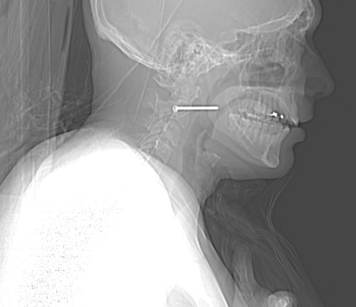
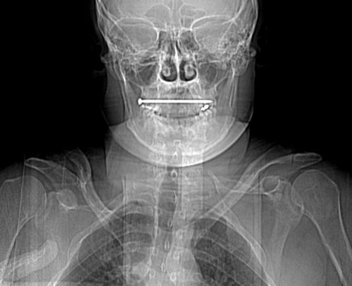
We’ve made sure that our victim of the nail gun to the neck did not need an emergent operation. Vitals are stable, there’s no uncontrolled hemorrhage, and the patient is neurologically intact. We’ve imaged him using CT angiography, and the nail does not appear to have injured any vital structures.

How do we get it out of there? There are two things that need to be considered: where and how.

Rule of thumb: If a foreign body is located anywhere near vital structures, take it out in the OR, no matter how good you think the imaging is. It may be tempting to just pull it out in the ED, but resist! CT scans look so good, and they are so detailed, but they are not perfect. The ED does not have the equipment, personnel, or lighting necessary if something goes awry.

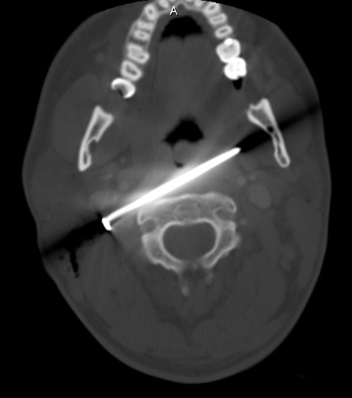
Rule of thumb: Use all information available to plan the removal procedure. In this case, the head of the nail is to the patient’s right. Therefore, it must be removed from the right side. The CT shows that the nail passes very close to the posterior pharynx, so it will need to be evaluated during the procedure.

This patient was taken to the operating room. During the intubation, direct laryngoscopy was carried out to carefully inspect the entire pharynx and larynx. No evidence of penetration was seen. The entire neck, face, and upper chest were prepped and draped (I like to go overboard in trauma cases; you never know what is going to happen). Fluoroscopy was available.

The classic operation would have been to make an incision along the sternocleidomastoid on the right side. The nail head would be approached directly. Since long, thin objects can be notoriously difficult to locate, fluoro can be very helpful. The exact position with respect to the carotid and jugular can be ascertained. Then the nail head is grasped with a clamp, and the nail gently pulled out along its axis. A nice, long wait for any evidence of bleeding should occur. The area can then be irrigated and the incision closed. Skin antibiotics can be given postop, but only one dose at most.

Having said that, I opted for a different approach based on an old blog post here. Tune in on Wednesday to see what I really did.

Print Friendly, PDF & Email

Nail In The Neck: Part 2

This case involves an accidental nail gun injury to the neck. The patient is hemodynamically stable, neurologically intact, the airway is patent and not threatened, and there is no apparent hematoma. There is a small puncture near the sternocleidomastoid muscle on the right, fairly high on the neck. The nail is not palpable on either side. And the patient only complains of a little discomfort when he swallows.

What to do? First, the patient has passed all the initial decision points that would send us straight to the OR (ABC problems in ATLS jargon). But, per physical exam and initial imaging, the nail must obviously come out. We just have to figure out what we need to know before we take it out, and determine the best way to retrieve it.

Given the patient’s stability, additional imaging will be helpful. Views in different planes, and details of what the nail might have passed through will be invaluable. The recommended study is a CT angio of the neck. This will give good information about nearby structures and the vasculature. And software reconstructions will provide good 2D/3D information for removal planning. Here’s a lateral view.

image

The nail is located in front of the body of C2. It appears to be high enough to be near the pharynx, but well above trachea and esophagus. You can also see that the nail entered a little posteriorly, and travels right to left and forward.

Here’s a representative CT slice.

image

The nail enters behind the carotids (just above the bifurcation) and IJ on the right, and ends anterior to them on the left. It passes very close to the posterior pharynx. So neurovascular structures are intact, and the aerodigestive tract is a maybe (back of the pharynx).

Obviously, this thing has to come out. The question is, how to do it? For you surgeons out there, tell me your choice of approach, incision, and instrumentation. Tweet or leave comments! Answers on Monday.

Print Friendly, PDF & Email

What Would You Do? Nail In The Neck

Here’s a very interesting case for you. A construction worker was carrying an object inside a building WHILE HOLDING HIS NAIL GUN! As he passed through the door, his elbow hit the frame and he brushed his neck with the business end of the gun. Guess what happened?

He experienced sharp pain, then noted pain every time he swallowed. He checked himself out in the mirror, and there was a small puncture wound in the right side of his neck. He presented to his local ED, with the complaints just noted. He was hemodynamically stable and neurologically intact. His airway was patent, and he had minimal pain. The following image was obtained. The nail measures about 6cm in length.

Let me know (by Twitter or comments) what additional information you think you need. I’ll discuss that in my next post. Then we’ll figure out what to do about this problem.

Print Friendly, PDF & Email

What Is: A Rubber Bullet?

The protests in cities across the country continue. Many are peaceful, but not all. In some cases, police have resorted to “non-lethal” weapons to control and disperse crowds.

Although these weapons are called non-lethal, that’s not entirely true. The projectiles, gases, and powders that are being used all have some degree of morbidity and mortality. They are certainly less so than traditional projectiles (bullets), but serious and fatal injures can and do occur.

One item that is talked about in the news is the rubber bullet. What are these, exactly? The generic term is a “kinetic impact projectile” (KIP). It encompasses a variety of objects that are not designed to penetrate flesh like a regular bullet. They can be bullets, beanbags, sponges, pellets, and other odds and ends.

And the so-called “rubber bullet” isn’t even necessarily made of rubber. It can be plastic, metal, rubber, or other substances.

There is very little published data on injuries caused by KIPs. Because of their odd shapes, they tend to tumble when they are fired. This decreases aiming accuracy substantially when the target is distant. They are designed to be aimed at the lower extremities. However, if the aim is too high or the round is fired at close range, it can be lethal.

Here are some typical injuries that have been descirbed:

  • Subdural and intraparenchymal hematomas
  • Skull and facial fractures
  • Eye injuries leading to blindness (this happened to a photographer in Minneapolis last week)
  • Rib fractures and pulmonary contusions
  • Spleen laceration
  • Blunt intestinal injury

Here’s a video from one of the manufacturers that shows the amount of target deformation caused by a sponge tipped bullet. Very impressive.

YouTube player

(Tumblr viewers please click here to view video)

Bottom line: Although they sound relatively innocuous, kinetic impact projectiles of any kind are far from it. If you are called to treat a patient who has been shot with one, be sure to do a very thorough evaluation. Most head, neck, and torso injuries should undergo CT scanning to delineate deep or occult injuries in detail. In-hospital observation of torso injuries is warranted, as well as a good tertiary exam.

Print Friendly, PDF & Email

What The Heck? CT Imaging Problem: The Answer

I received some good guesses about this image yesterday, but no one got the right answer.

The patient had sustained blunt trauma and was undergoing CT imaging. The scout for the abdominal CT showed some kind of weird debris that interfered with the image, but when we uncovered and looked at the patient, nothing was visible:

What the heck? If you look carefully at the left side of the image, you can see that the “debris field” is on the surface of the patient. We can’t see in 3-D on images, but the difference in appearance on the left and right sides looks like it this stuff is wrapping around the patient.

She was brought in by EMS with a warming blanket in place. On closer inspection, this was a thin, disposable blanket that heats up when removed from an airtight plastic pouch. These blankets contain thin pockets of a mineral mixture that looks like gravel. When exposed to air it heats up.

But on CT it looks like bone density material! When we looked at the patient, we were just lifting off the blanket that contained the offending material. Hence, we couldn’t find it.

Here’s a picture of one of these products. Note the six mineral pouches embedded in it., Don’t let this happen to you!


Print Friendly, PDF & Email