Category Archives: Thorax

Early Vs Delayed Thoracic Endovascular Repair

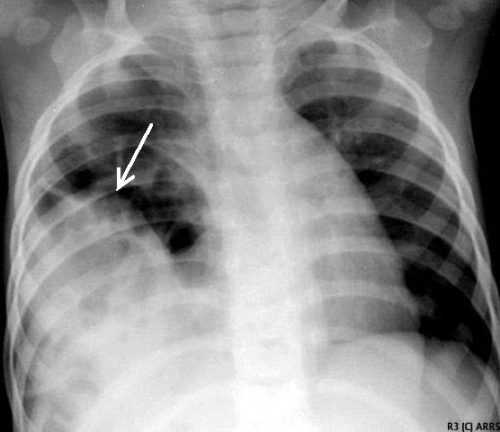
Back in the day, the only way to fix a broken thoracic aorta was via left thoracotomy.  This was a big procedure, with the possibility of several major complications, with postop paraplegia being one of them. At the time, there was a debate about whether the procedure should be done immediately versus waiting until the patient was well-resuscitated. The concern was that death was nearly certain if the aortic lesion progressed.

We learned that temporizing with strict blood pressure control worked wonders at protecting the patient. Although many of these injuries were managed within hours, a growing number were delayed by a few days to improve outcomes.

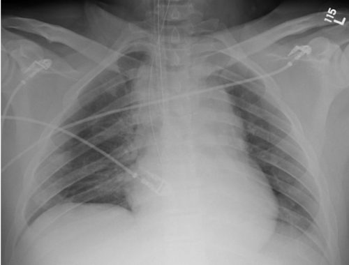
Nowadays, thoracic endovascular aortic repair (TEVAR) is routine and much less morbid than the open procedure. However, the same question arises: do it early or wait a while? Interestingly, not one but two analyses have been published on this very topic in the last four months!

The first is from an international research group that searched the usual databases and initially found 921 records. They included only clinical trials or cohort studies with ten or more adult patients that could be stratified as early (within 24 hours) or late (after 24 hours) intervention. After applying these criteria, only seven studies remained for analysis.

There were 3,757 patients with early repairs, compared to 1,238 undergoing late repair. The presenting demographics and injury grades were similar in each group. However, the short-term mortality was significantly higher (1.9x) in the early TEVAR group. Additionally, ICU length of stay was significantly longer (3 days) in the late TEVAR group.

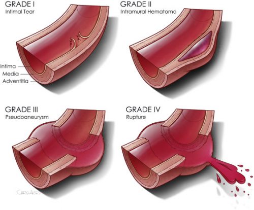
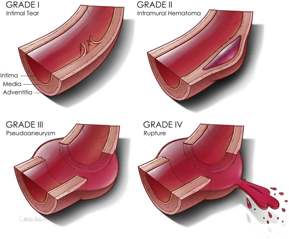
The second paper was presented as a quick-shot at last year’s AAST meeting. It is from a group of researchers from our big Boston trauma centers and the Netherlands. They used four years of data from the TQIP database, giving them extra information unavailable in the first study. They specifically looked at patients with grade II or III injuries. Here is the grading scale:

Here are the factoids:

  • A total of 1,339 patients were studied, with about three-quarters in the early TEVAR group
  • Median time to TEVAR was 4 hours in the early group and 65 hours in the late group
  • Patients in the early group were significantly less likely to have brain or liver injuries
  • ISS was similar in both groups
  • The early TEVAR group had significantly higher in-hospital mortality (16% vs. 5%), significantly higher risk of ARDS (7.6% vs. 2.1%), but significantly shorter ICU stay (7 vs 10 days)
  • When patients who died within the first 24 hours were excluded, the in-hospital mortality remained significantly higher, and the ICU and hospital lengths of stay were significantly shorter

Bottom line: Some society guidelines began recommending delayed TEVAR in 2015. This study did not detect any trend toward this, however. Using different methods and databases, these two studies identified nearly identical mortality and ICU trends in large groups of patients. The mortality trends do not appear to be related to injury grade, overall injury severity, or the presence of head injury. 

Taken together, this suggests that we need to rethink the timing of TEVAR in patients with grade II or III injuries. The best timing still needs to be defined, but it appears to be beyond 24 hours. Centers performing this procedure should review their results and consider extending procedure timing as additional research is done to define the ideal time interval.

References:

  1. Early Versus Delayed Thoracic Endovascular Aortic Repair for Blunt Traumatic Aortic Injury: A Systematic Review and Meta-Analysis. Cureus. 2023 Jun 28;15(6):e41078. doi: 10.7759/cureus.41078. PMID: 37519486; PMCID: PMC10375940.
  2. Early Versus Delayed Thoracic Endovascular Aortic Repair for
    Blunt Thoracic Aortic Injury: A Propensity Score-Matched Analysis. Ann Surg 278:e848-e854, 2023.


Best Of AAST #3: When To Place A Chest Tube For Hemothorax

There is an art to deciding when to place a  chest tube for either hemothorax or pneumothorax. For the most part, the trauma professional examines the imaging and then uses some unknown internal metric to declare that it is “too big.” Then it’s time to insert some type of chest drain.

There have been attempts over the years to make this decision more quantitative. One of the better-known ones is the 2-cm rule for pneumothorax. If the distance from the chest wall to the lung on the chest x-ray is >2cm, it is “too big.”

But what about hemothorax? The Medical College of Wisconsin trauma group performed a retrospective review of 391 patient charts to test a new 300cc rule defining when a hemothorax is “too big.” This guideline was implemented in 2018-2019, and patients presenting before implementation were compared to those arriving after.

The 300cc threshold is determined by using Mergo’s formula for calculating the volume of a square prism. Obviously, this requires a CT scan for calculation, so patients who had a tube placed before scanning or did not have one were excluded. They were also excluded from the study if their pneumothorax met the 2-cm rule. The authors studied how many patients could be observed, how many needed tube drainage, observation failure, and later need for a VATS procedure or thoracotomy.

Here are the factoids:

  • About 60% of the study group was admitted after the new criteria were implemented, and both groups were demographically similar
  • After implementation, the number of patients that were just observed increased significantly from 52% to 71%
  • Of course, this means that the number of chest tubes inserted was significantly less (42% vs. 61%)
  • There was no difference in observation failure (delayed placement of a tube), 18% vs. 24%
  • There were also no differences in pulmonary complications, 30-day readmissions, or 30-day mortality
  • The average ICU and hospital length of stays were significantly shorter as well

The authors concluded that implementing their 300cc guidelines correlated with decreased length of stay and no increase in failure or complication rates.

Bottom line: Although this is a relatively small series, the differences between the groups quickly achieved significance. There are three major questions that I have. First, how was the 300cc threshold arrived at? Was this borne of clinical judgment, or did some previous work suggest it?

My next question has to deal with the accuracy of the volume calculation. Mergo’s formula was used to determine the volume of a rectangular solid. As we all know, hemothoraces and pneumothoraces are not cubes. They can be very irregular and influenced by patient position. However, I did find a paper from the University of Florida that found the correlation coefficient between the volume calculated by Mergo’s formula vs. using 3-D software estimation was 0.9, which is excellent. So this approximation appears to be a very good one.

Finally, using the 300cc rule is predicated on getting a CT scan. Does every patient need a chest CT? Part of the resuscitation process for major trauma involves obtaining a chest X-ray. The obviously large hemothorax can justify inserting a chest tube at that point. But the reality is that most of these patients do go on to chest CT, so this is a minor change in practice for most. 

Although I love to see confirmatory studies before practice changes, this one study can lead us to change our practice guidelines now. It is a relatively minor one and will allow us to avoid placement of a few more chest tubes and to shave off a few days of hospital stay. The logical follow-up study for the authors is to extend the post-discharge window for complications to 60 or 90 days to ensure that delayed procedures were not required in the observation group.

References:

  • Implementing the 300-cc rule safely decreases chest tube placement in traumatic hemothorax. AAST 2023 Plenary paper #22.
  • New formula for quantification of pleural effusions from computed tomography. J Thorac Imaging. 1999 Apr;14(2):122-5. 


Delayed Presentation Of Right Diaphragm Injury

Diaphragm injury from blunt trauma is uncommon, occurring in only a few percent of patients after high-energy mechanisms. They usually occur on the left side and are more frequently seen after t-bone type car crashes and in pedestrians struck by a car.

Blunt diaphragm injury on the right side is very unusual. Even so, it is more easily detected due to obvious displacement of the liver that can be seen on chest x-ray. Blunt injuries on the right side usually result in a large rent in the central tendon or detachment of the diaphragm from the chest wall. This allows the liver to herniate into the chest, and the chest x-ray finding is not subtle.

This image shows an acute herniation of the liver through the diaphragm. Due to the size of the liver, only part of it can typically fit through the rent. Radiologists call this the “cottage loaf” sign. Why? Here’s the bakery item it is named after. Get it now?

Thankfully, most of these injuries are identified in the acute setting. They must be addressed surgically because, if left untreated, more and more of the liver will slowly move into the chest resulting in respiratory problems in the long run.

Acute management usually consists of laparotomy to address both the diaphragm tear and any other associated intra-abdominal injuries. The liver should be reduced by sliding a hand next to it laterally into the chest cavity and pushing the dome downwards. The right triangular ligaments should be taken down (if they are not already destroyed) to mobilize the organ better so the diaphragm laceration can be closed. This is typically accomplished with some type of large (size 0) permanent suture. A chest tube will be needed to evacuate the iatrogenic pneumothorax created by opening the abdomen.

Chronic right diaphragm injuries are a different animal entirely. There is no longer any need to evaluate for intra-abdominal injury, so the procedure is usually performed through the chest. For smaller injuries, thoracoscopic procedures have been described that push the liver downwards and then either suture the diaphragm primarily or (more likely) incorporate a piece of mesh.

Larger injury requires conversion to an open procedure so more muscle power can be used to push the liver downwards to facilitate the repair. However, do not underestimate the adhesions that will be present between diaphragm and liver (and possibly the lung) in long-standing injuries. It may take some time to dissect them away. Rarely, a laparotomy (or laparoscopy) may be needed to assist for very large and complex injuries.

References:

  • Management of Delayed Presentation of a Right-Side Traumatic
    Diaphragmatic Rupture. World J Surg 36:260-265, 2012.
  • Delayed Discovery of Diaphragmatic Injury After Blunt Trauma:
    Report of Three Cases. Surg Today 35:407-410, 2005.

Flying Or Diving After Traumatic Pneumothorax: Part 1

Today, I’m dusting off an old post on flying and diving after pneumothorax. This shows the thinking up until a few years ago. In my next post, I’ll write about a more recent paper that suggests that we can shorten the “no-fly” time considerably.

Hint: no changes to the diving recommendations. One pneumothorax is likely to ground you forever. (pardon the pun)

Patients who have sustained a traumatic pneumothorax occasionally ask how soon they can fly in an airplane or scuba dive after they are discharged. What’s the right answer?

The basic problem concerns Boyle’s Law (remember that from high school?). The volume of a gas varies inversely with the barometric pressure. So the lower the pressure, the larger the volume of gas becomes. Most of us hang out close to sea level, so this is not an issue. But for flyers or divers, it may be.

Flying

Helicopters typically fly only one to two thousand feet above the ground, so the air pressure is about the same as standing on the earth. However, flying in a commercial airliner is different. Even though the aircraft may cruise at 30,000+ feet, the inside of the cabin remains considerably lower though not at sea level. Typically, the cabin altitude goes up to about 8,000 to 9,000 feet. Using Boyle’s law, any volume of gas (say, a pneumothorax in your chest) will increase by about a third on a commercial flight.

The physiologic effect of this increase depends upon the patient. They may never know anything is happening if they are young and fit. But if they are elderly and/or have a limited pulmonary reserve, it may compromise enough lung function to make them symptomatic. And having a medical problem in an aluminum tube at 30,000 feet is never good.

Commercial guidelines for travel after pneumothorax range from 2-6 weeks. The Aerospace Medical Association published guidelines that state that 2-3 weeks is acceptable. The Orlando Regional Medical Center reviewed the literature and devised a practice guideline with a single Level 2 recommendation that commercial air travel is safe 2 weeks after resolution of the pneumothorax, an that a chest x-ray should be obtained immediately before travel to confirm resolution.

Diving

Diving would seem to be pretty safe, right? Any pneumothorax would just shrink while the diver was at depth, then re-expand to the original size when he or she surfaces, right?

Not so fast. You are forgetting why the pneumothorax was there in the first place. The lung was injured, most likely via tearing it, penetration by something sharp, or popping a bleb. If the injured area has not completely healed, then air may begin to escape through it again. And since the air used in scuba diving is delivered under pressure, this could result in a tension pneumothorax.  This is disastrous underwater!

Most injuries leading to pneumothorax heal completely. However, if there are bone spicules stuck in the lung or more complicated parenchymal injuries from penetrating injury, they may never completely heal. This makes the diver susceptible to a tension pneumothorax anytime they use their regulator.

Bottom line: Most patients can safely travel on commercial aircraft 2 weeks after resolution of pneumothorax. Ideally, a chest xray should be obtained shortly before travel to confirm that it is gone. Helicopter travel is okay at any time, since they typically fly at 1,500 feet or less.

Divers should see a physician trained in dive medicine to evaluate their injury and imaging prior to making another dive.

Tomorrow: new info on flying after pneumothorax

References:

  • Divers Alert Network – Pneumothorax – click to download
  • Practice Guideline, Orlando Regional Medical Center. Air travel following traumatic pneumothorax. October 2009.
  • Medical Guidelines for Airline Travel, 2nd edition. Aerospace Medical Association. Aviation, Space, and Environmental Medicine 74(5) Section II Supplement, May 2003.

Best Of AAST 2022 #9: Management Of Low Grade Blunt Thoracic Aortic Injury

It has been interesting to watch the evolution of the treatment of blunt thoracic aortic injury (BTAI). In my training, patients with an abnormal mediastinal contour on chest x-ray were whisked away to interventional radiology for a thoracic aortic angiogram. Yes, there was no such thing as a CT scan for anything but the head! Here’s a sample chest x-ray:

I wrote a post 12 years ago describing the various findings of a blunt aortic injury, many of which are present above. You can the post by clicking here.

In the 1-2% where the angio was positive (yes, you read that right; we did a lot of negative angiograms) the patient was then whisked off to the OR for an open aortic graft via thoracotomy. The advent of CT scans of the torso and improving resolutions allowed us to be more selective with angiography. And finally as CT angiography matured, aortic angiography became a thing of the past.

Then came TEVAR about 20 years ago. There were some growing pains as we refined the technology, but now this endovascular procedure is the standard of care for most aortic injuries. While there was also a place for nonoperative management, it was really just maintaining a reasonably low blood pressure to protect the vessel while getting the patient into good enough condition to tolerate anoperation.

Now we are beginning to slice and dice treatment based on grade. Many centers have recognized that an intimal injury (Grade I) is only a very minor disruption to the inside of the vessel and does not make it more susceptible to rupture. Grade II management has been less clear. Here is a diagram of the various grades.

It makes sense that an invasive procedure may be less helpful for injuries that do not disrupt the layer of the vessel that provides its strength. But remember, common sense isn’t always the truth.

The group at Dell Seton Hall reviewed all patients with low grade BTAI (Grades I and II) in the Aortic Trauma Foundation Registry for a six year period. Their hypothesis was that these injuries could be successfully treated with medical management alone. They reviewed the data for mortality, complications, vent days, and lengths of stay.

Here are the factoids:

  • A total of 880 patients were enrolled and 274 had low grade injuries; 5 were then excluded when their lesion progressed and they underwent TEVAR
  • Of the 269 remaining patients, 81% were treated with medical management (81% Grade I, 19% Grade II) and the remainder with TEVAR (20% Grade I, 80% Grade II)
  • Rates of thoracotomy, craniectomy, and sternotomy were the same in both groups, but TEVAR patients were more likely to have a laparotomy (31% vs 15%)
  • Mortality was significantly higher in the TEVAR group (18% vs 8%) but the mortality from the aorta was not quite significant (4% vs 0.5%)
  • Complications (DVT and ARDS) were also significantly higher in the TEVAR group
  • Vent days and lengths of stay were equivalent

The authors concluded that medical management alone is safe and appropriate, with a lower mortality and decreased complications compared to routine TEVAR.

Bottom line: Hmm, color me skeptical. Remember, this is a registry study, so information tends to be limited outside the usual demographics and data points that are very pertinent to the purpose of the registry. The most important concept is that the patients in each group must be identical in every way except for the intervention.

We can try to make them as identical as possible by matching pairs or subgroups of patients. But the biggest problem is that the number of patient characteristics that might be important to match may not be available for analysis in the database.

When the patients were initially treated at contributing centers, there were no specific rules that the individual surgeons had to follow to decide between medical management and TEVAR. They could pick and choose based on their own experience. Could the surgeon have recognized some patients as higher risk and opted for TEVAR to make sure the aorta would not become an issue in conjunction with their other injuries? And unfortunately, perhaps that higher risk issue is what ultimately killed them and not the aorta. It’s basically a form of unhealthy user bias.

This abstract is an interesting tidbit that should push us to question whether medical management is better, at least in some subsets of low grade aortic injury patients. Then someone can perform a more robust study to confirm or refute its safety. Unfortunately, this may never happen due to the low incidence of this injury. It took six years to accumulate only 269 eligible patients in this registry!

Here are my questions for the authors and presenter:

  • What data points are actually in the registry? Specifically, is there fine detail about the other injuries the patient had? Could these have contributed to TEVAR mortality, or the selection of the patient for TEVAR to try to reduce the perceived mortality risk?
  • When you stated that there was “no difference in demographics or mechanism of injury” what were these, exactly?
  • What did patients actually die from in the Grade I and Grade II groups? Be more specific than “aortic-related” or not.
  • Do you have any worries about the five patients whose lesions progressed? When should patients be re-scanned to identify those who might benefit more from TEVAR?

This is a thought provoking abstract, and I am very interesting in hearing what the next steps should be.

Reference: MEDICAL MANAGEMENT IS THE TREATMENT OF CHOICE FOR LOW GRADE BLUNT THORACIC AORTIC INJURIES. Plenary paper #45, AAST 2022.