Category Archives: Solid organ

Surveillance For Splenic Pseudoaneurysm After Injury

When it comes to repeat CT scanning after splenic injury, there are believers and there are non-believers. In my experience, the majority of centers in the US are non-believers. However, there is a new paper in press that attempts to convince us that more should become believers.

I think the biggest lesson to be learned from this paper is that WE SHOULD READ THE ENTIRE PAPER before drawing conclusions. I have said this in the past and I will say it again. In this case, not only did I read the entire paper, but I had to dig deep into the references it cited as well.

Nonoperative management of splenic injuries has a very high success rate if done properly. Some papers claim this can be up to 93%, which parallels my experience. This success rate involves excluding unstable patients (they need to be in the operating room) and planned use of angioembolization in select patients. Over the years we have found that we need to do less and less in the management of solid organ injury patients:

  • No bedrest
  • No starvation (NPO status)
  • No serial blood draws
  • No repeat CT scan
  • Few limitations on activity after discharge

For an example of a practice guideline that demonstrates that less is more, use the download link at the end of this post.

But back to the question about repeat CT scanning before discharge. Why do we need to do this? The usual reason is that “we want to find delayed pseudoaneurysms.” And why is that important? “It might bleed!”

Really? Let’s look into that through the lens of this new paper by the group at the University of Cincinnati. They performed a retrospective study of their experience with patients who had sustained blunt splenic injury during a recent three-year period. They were interested in how many underwent splenectomy or splenorrhaphy, who had repeat CT imaging, who went to interventional radiology (IR) and when, and which ones were found to have pseudoaneurysms and what was done about it.

Here are the factoids:

  • There were 539 patients who met inclusion criteria, with an average ISS of 24
  • Of these, 46 died during their hospital stay (none from their splenic injury)
  • Focusing on the 248 patients with higher grade injuries (III-V), 125 (50%) underwent emergent or delayed splenectomy. Early vs late operation was not broken out, but this is a startlingly high number!
  • Of the higher grade injured patients who kept their spleens, 97% underwent repeat CT around day 5
  • Delayed pseudoaneurysms were detected in the following patients:
    • Grade III: 10 of 88 patients (11%). Then 8 of those 10 went to IR, and 5  of 10 had splenectomy!
    • Grade IV: 7 of 24 (29%).  Then 8 of the 7 (error in the paper?) went to IR and 3 of 7 had splenectomy!
    • Grade V: 2 of 5 (40%). Both of these patients went to IR and somehow kept their spleens.

The authors conclude that routine followup CT imaging identifies splenic pseudoaneurysms allowing for interventions to minimize delayed complications.

Bottom line: Whoa! There’s a lot going on here. My first observation is that this center does a lot of splenectomies! Of the 539 patients (all comers) who were included in the study, 129 (24%)  lost their spleens. If grade I-II injuries are excluded that percent rises to 50%!

Only eight splenectomies were performed after the repeat CT. This would imply that there were either a lot of unstable patients with splenic injury, the institutional indications for this procedure arbitrarily include grade, or there is a lot of variability in the decision to perform it.

I think there are really two questions to answer here. 

  1. Does delayed splenic pseudoaneurysm occur? The answer is yes. There are a few studies (performed by believers) that demonstrate new pseudoaneurysms after repeat CT. I’m convinced.
  2. Do we care? The real question is, do these pseudoaneurysms cause harm? The fear is that they might explode at some point after patient discharge and cause a major problem.

Papers written by the believers cite a number of old studies and give numbers between 2% and 27% for incidence of delayed hemorrhage. Well, I tracked down all of these papers, including the ones they cited. And it doesn’t add up.

  • One paper from a believer institution found no delayed bleeds.
  • Several papers were for pediatric patients, whose spleens don’t behave like adult ones. They found one case after discharge in one out of 276 patients across three studies.
  • Of 76 adolescents, none encountered delayed bleeds

Many of the papers cited regarding bleeding complications are very old. CT scanners had less resolution, and in many papers, IR was not even a consideration. 

So here’s my take. Yes, delayed pseudoaneurysms occur. In children we don’t care. They almost never cause a problem. But in adults, they can and do cause issues and should be embolized shortly after the initial scan. 

Once embolized, the ones seen on that initial scan are effectively neutralized and do not need a repeat scan. The small ones that might pop up later may very well be part of the healing process. And they may not even occur if angioembolization is done early. It seems unlikely that anything further is needed.

But remember, clinical judgement trumps all. If your patient starts complaining of new abdominal symptoms while in the hospital or after discharge, get a prompt CT scan to rule out any developing complications.

Sample solid organ injury protocol: click here

Reference: Delayed splenic pseudoaneurysm identification with surveillance imaging. J Trauma Acute Care Surg. 2022 Mar 22. doi: 10.1097/TA.0000000000003615. Epub ahead of print. PMID: 35319540.


Print Friendly, PDF & Email

Does Time To Angioembolization Make A Difference?

Angiography and angioembolization (AE) have helped trauma professionals stop pesky bleeding from the pelvis, solid organs, and other hard to reach places for decades. The American College of Surgeons has recognized its importance and even has a specific criterion for interventional radiologist response times.

Many centers have found this 30-minute response for radiologist arrival to be onerous. So, of course, someone decided to look at the data to see if it was really warranted. The group at the University of Arizona at Tucson did a big data dive using the Trauma Quality Improvement Program database to try to tease out the consequences of prompt vs delayed access to AE in blunt trauma patients.

Four years of TQIP data were analyzed. The authors focused on the records of patients who underwent AE within 4 hours of arrival for blunt injury to liver, spleen, or kidney. They excluded transfers in, burn patients, and those who underwent an operation prior to AE. The 4 hour AE period was subdivided into hourly segments and outcomes were examined for each.

Here are the factoids:

  • Out of over a million records, 924 met study inclusion criteria
  • Patients were relatively young with a mean age of 44, and seriously injured with a median ISS of 29
  • Spleen injuries were most common (64%), followed by liver (50%), and kidney (27%)
  • Overall 24 hour mortality was 5% and in-hospital mortality was 15%
  • The 24 hour mortality significantly increased as each hour passed until AE, from 2% to 24%. 
  • On multivariate analysis, in-hospital mortality showed the same hourly increase
  • No differences in the amount of blood, plasma, or platelets given were noted in any of the groups
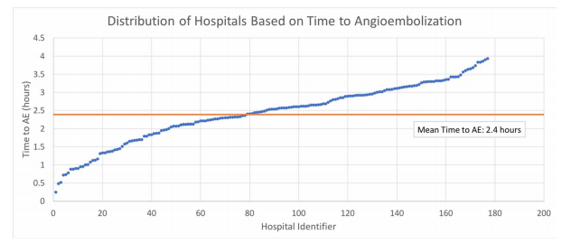
  • Average time to AE correlated with trauma center AE volume, with 1.6 hours at centers doing more than 14 cases per year to 2.7 hours at those doing less than 9 cases

The authors concluded that delayed AE for solid organ injury resulted in increased mortality without any difference in transfused blood products. They encouraged centers to ensure rapid access to this vital procedure.

Bottom line: This is an important paper. Other research on angiography in blunt trauma has not shown the survival differences that we see here. But they did not focus on patients who underwent actual embolization. This one shows that time to AE is very important in those patients who really need it.

It appears that the most important factor is door to angiography suite time. There are many factors involved, though. First, the specific injury must be recognized by the clinicians. This involves access to CT, timeliness of radiologist report, decision making by the trauma professional, and timeliness of response by the interventional radiologist and the IR team.

Most centers focus only on that 30-minute response time required of the interventionalist. But as you can see, there are many more moving parts. I would urge all centers to look at their door to AE time and compare to what was found in this study. If you find that it is taking more than an hour and a half, it’s probably time for a PI project to tighten it up. This is particularly important in centers with low AE volume.

Reference: Angioembolization in intra-abdominal solid organ injury: Does delay in angioembolization affect outcomes? J Trauma 89(4):723-729, 2020.

Print Friendly, PDF & Email

VTE Prophylaxis After Solid Organ Injury

Venous thromboembolism (VTE)  is a common potential complication after traumatic injury. But typically, injury is associated with bleeding, so the trauma professional has to strike a balance between preventing bleeding and preventing clots.

Solid organ injury (liver and spleen, typically) is a common diagnosis after blunt trauma. Most trauma centers have protocols for VTE prophylaxis which apply to patients with those injuries. Older literature that I wrote about eight years divided the time frames for prophylaxis into early (within 3 days), late (greater than 3 days), and none. The authors of that article found that there was no association with untoward bleeding in the early group. And interestingly, there seemed to be less in that group. Unfortunately, the selection of the groups was biased, and the early VTE prophylaxis group had less severe injuries.

The surgery group at the Massachusetts General Hospital tried to clarify current practice by performing a deep dive into the Trauma Quality Improvement Program database. They searched the database to identify patients with “isolated” liver, spleen, kidney, and pancreas injury. They did this by excluding TBI, femur and pelvic fractures, spinal cord injury, and penetrating trauma. They also excluded patients with other other severe injuries with an abbreviated injury scale score of 3 or more.

The authors stratified patients into three groups: early VTE prophylaxis receiving the drug within 48 hours of arrival, intermediate within 48-72 hours, and late after 72 hours.

Here are the factoids:

  • A total of 3,223 patients met inclusion criteria
  • Prophylaxis was classified as early in 57%, intermediate in 22%, and late in 21%
  • About 3/4 received low molecular weight heparin and the remainder received unfractionated heparin
  • Late prophylaxis was associated with a 3x increase in both VTE and pulmonary embolism (PE)
  • Intermediate prophylaxis patient had a 2x increase in VTE but no increase in PE
  • Early prophylaxis showed a 2x increase in bleeding complications, especially in those with diabetes (?), spleen, and high-grade liver injury
  • A total of 60 of the 1,832 patients in the early group had bleeding events: 39 failed nonop mangement and were taken to OR, 8 underwent angioembolization, and 21 received blood transfusions

The authors concluded that early prophylaxis should be considered in patients who do not fall out as higher risk (spleen, high-grade liver, diabetics).

Bottom line: This retrospective study is probably as good as it’s going to get from a data quality standpoint. It’s larger than any single-institution series will ever be, although it suffers from the usual things most large database studies do. 

But it does show us strong associations with DVT and PE as the consequences of waiting to start VTE prophylaxis beyond 48 hours. The caveat is to be careful in certain patients, most notably diabetics and those with liver and spleen injuries, as they are at higher risk to develop complications leading to the OR or interventional radiology suite. 

I urge all of you to re-examine your VTE prophylaxis guideline and modify it to start your drug of choice as early as possible given the cautions for patients with spleen and high-grade liver injuries. The diabetes thing, well, that’s a mystery to me and I will wait for further confirmation to break those patients out separately.

If you are interested, you can see the Regions Hospital trauma program VTE guideline by clicking here.

References:

  • Thromboembolic prophylaxis with low-molecular-weight heparin in patients with blunt solid abdominal organ injuries undergoing nonoperative management: current practice and outcomes. J Trauma 70(1): 141-147, 2011.
  • Timing of thromboprophylaxis in patients with blunt abdominal solid organ injuries undergoing nonoperative management. J Trauma pulish ahead of print, October 12, 2020, doi: 10.1097/TA.0000000000002972
Print Friendly, PDF & Email

Does Time To Interventional Radiography Make a Difference In Solid Organ Injury?

Solid organ injury is one of the more common manifestations of blunt abdominal trauma. Most trauma centers have some sort of practice guideline for managing these injuries. Frequently, interventional radiology (IR) and angioembolization (AE) are part of this algorithm, especially when active bleeding is noted on CT scan.

So it makes sense that getting to IR in a timely manner would serve to stop the bleeding sooner and help the patient. But in most hospitals, interventional radiology is not in-house 24/7. Calls after hours require mobilization of a call team, which may be costly and take time.

For this reason, it is important to know if rapid access to angioembolization makes sense. Couldn’t the patient just wait until the start of business the next morning when the IR team normally arrives?

The group at the University of Arizona at Tucson tackled this problem. They performed a 4-year retrospective review of the TQIP database. They included all adult patients who underwent AE within four hours of admission. Outcome measures were 24-hour mortality, blood product usage, and in-hospital mortality.

Here are the factoids:

  • Out of over a million records in the database, only 924 met the inclusion criteria
  • Mean time to AE was 2 hours and 22 minutes, with 92% of patients getting this procedure more than an hour after arrival
  • Average 24-hour mortality was 5%. Mortality by hours to AE was as follows:
    • Within 1 hour: 2.6%
    • Within 2 hours: 3.6%
    • Within 3 hours: 4.0%
    • Within 4 hours: 8.8%
  • There was no difference in the use of blood products

The authors concluded that delayed angioembolization for solid organ injury is associated with increased mortality but no increase in blood product usage. They recommend that improving time to AE is a worthy performance improvement project.

Bottom line: This study has the usual limitations of a retrospective database review. But it is really the only way to obtain the range of data needed for the analysis. 

The results seem straightforward: early angioembolization saves lives. What puzzles me is that these patients should be bleeding from their solid organ injury. Yet longer delays did not result in the use of more blood products.

There are two possibilities for this: there are other important factors that were not accounted for, or the sample size was too small to identify a difference. As we know, there are huge variations in how clinicians choose to administer blood products. This could easily account for the apparent similarities between products given at various time intervals to AE.

My advice? Act like your patient is bleeding to death. If the CT scan indicates that they have active extravasation, they actually are. If a parenchymal pseudoaneurysm is present, they are about to. So call in your IR team immediately! Minutes count!

Reference: Angioembolization in intra-abdominal solid organ injury:
Does delay in angioembolization affect outcomes?  J Trauma 89(4):723-729, 2020.

Print Friendly, PDF & Email

Best of AAST #12: Embolization Of Splenic Pseudoaneurysm

The management of blunt spleen injury has evolved significant over the time I’ve been in practice. Initially, the usual formula was:

Spleen injury = splenectomy

This began to change in the late 1980’s, and beginning in the early 90’s nonoperative management became the rage. We spent the next 10-15 years tweaking the details, gradually reducing bed rest and NPO times, and increasing the success rate through smart patient selection and discovering new adjuncts.

One of these adjuncts was angiography with embolization. The ShockTrauma Center in Maryland was an early adopter and protocolized its use in patients with high-grade injuries.

But now, they are questioning the utility of this tool in certain patients: those with splenic pseudoaneurysms (PSA). They theorized that modern, high resolution CT identifies relatively unimportant pseudoaneurysms. They conducted a 5-year retrospective review of their experience.

Here are the factoids:

  • They identified 717 splenic injuries, of whom 155 were embolized but only 140 patients had adequate records and imaging for review
  • The majority of patients had high grade injury: 31% Grade 3, 61% Grade 4, 1% Grade 5
  • Extravasation was seen in 17% and PSA in 52%
  • About 44% of patients went to angiography within 6 hours, but the mean was 17 hours indicating quite a few outliers
  • Among the 73 patients with an initial PSA , a third of them did not have a detectable lesion during angiography
  • Patients who underwent embolization for PSA had a followup CT 48-72 hours afterwards, persistently perfused PSA were seen in 40% (!)
  • No patients with PSA who were only observed required delayed splenectomy

The authors conclude that a third of pseudoaneurysms may be clinically insignificant, and that 40% of them persist after embolization. They do not, however, offer any recommendations based on their data.

Here are my comments: This is an interesting study. My read of the abstract and slides would indicate that this group routinely sends all Grade 3 and 4 injuries to angio, and Grade 5 could go to either angio or OR. They take their good time going to interventional radiology (mean 17 hours from arrival), and get a routine followup CT 48-72 hours from hitting the door if they didn’t go to the OR.

If I were to play the devil’s advocate, I might think that interventional radiology was being de-emphasized for some reason. Was there some reluctance to send patients there, or limited availability? This might explain the long access times. And how are the radiologists not shutting down 40% of PSA that are seen?

I am intrigued by the study, but there are a lot more details needed to get some good takeaways from it.

Here are my questions for the presenter and authors:

  • Please explain why it takes so long to send patients to angiography. Less than half got there in less than 6 hours, and the mean of 17 hours means that many didn’t get there until the next day.
  • Does this small study have the statistical power to say that some PSA are benign? The groups are very small, and I would speculate that the group size needed to show significance is in the high hundreds.
  • What was the reason for splenectomy in the 2 patients who underwent embolization? Was it related to the pseudoaneurysm or something else?
  • How can you be sure that these PSA are insignificant? Frequently, pseudoaneurysms don’t explode for 7-10 days. Do you have any data on patients who returned to a hospital with delayed bleeding?
  • If you believe that many pseudoaneurysms are benign, how do you propose to manage the patients? Observe until they explode? Repeat a contrast CT scan, with the associated contrast and radiation re-dose? And how long would you wait to do this? What would your new protocol be?

I’ll be all ears on Friday when this abstract is presented live.

Print Friendly, PDF & Email