Category Archives: Resuscitation

How To Remember Those “Classes of Hemorrhage”

The Advanced Trauma Life Support course lists “classes of hemorrhage”, and various other sources list a similar classification for shock. I’ve not been able to pinpoint where these concepts came from, exactly. But I am sure of one thing: you will be tested on it at some point in your lifetime.

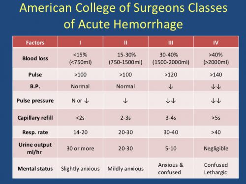
Here’s the table used by the ATLS course:

classes_of_shock

The question you will always be asked is:

What class of hemorrhage (or what % of blood volume loss) is the first to demonstrate systolic hypotension?

This is important because prehospital providers and those in the ED typically rely on systolic blood pressure to figure out if their patient is in trouble.

The answer is Class III, or 30-40%. But how do you remember the damn percentages?

multiscore-maxi1

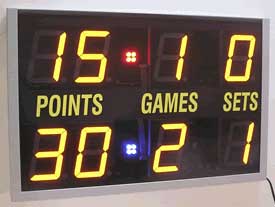
It’s easy! The numbers are all tennis scores. Here’s how to remember them:

Class I up to 15% Love – 15
Class II 15-30% 15 – 30
Class III 30-40 30 – 40
Class IV >40% Game (almost) over!

Bottom line: Never miss that question again!

Print Friendly, PDF & Email

ACS Trauma Abstract #6: Scanning Unstable Trauma Patients???

If you’ve read my stuff for very long, you know I frown on sending unstable patients anywhere but to the OR. Instability tends to get worse, and that always happens at inopportune locations like hallways, elevators, and CT scanners. Imagine my surprise when I noticed an abstract being presented at the Clinical Congress of the American College of Surgeons this week suggesting that it was okay to scan hemodynamically unstable patients before “definitive therapy.”

Here’s the title:

“Computed tomography in hemodynamically abnormal thoracoabdominal trauma safely enhances surgical triage”

The devil is in the details and the language. This group from USC included all patients who were hemodynamically abnormal on arrival to the trauma bay but who normalized to SBP > 90 during the resuscitation were included.  A total of 253 of these patients were reviewed over a 9 year period, and the usual variables were analyzed (mortality, complications, hospital, ICU, and vent days, etc).

Here are the factoids:

  • Of the 253 patients studied, 45 went to straight to OR and 208 were taken to CT
  • Injury severity was identical for the two groups
  • Lengths of stay and mortality were not different, but only p values were given
  • Patients taken to CT cleared their lactic acidosis faster (12 vs 5 hours), and used a bit less plasma and significantly less blood transfusions
  • The OR group underwent more procedures (31% vs 13%), although what these were and when they were performed is not listed

Bottom line: The title of this abstract is misleading, and may fool those who don’t read the rest of the abstract. It should read:

“Computed tomography in previously hemodynamically abnormal thoracoabdominal trauma safely enhances surgical triage”

Someone who just skims through this issue of the journal may get the idea that it’s okay to scan an unstable patient. The authors are not saying this at all. If you read the conclusion carefully, you can see that the patients had to be resuscitated to a SBP > 90 before they considered taking to scan. And they did that for the majority of these patients.

The real question is, why do the scanned patients clear their lactic acidosis faster, need less blood, and undergo fewer procedures? It appears that there is some bias or selection process in play. Otherwise, why not use the magic CT scanner to make them all better?

Reference: Computed tomography in hemodynamically abnormal thoracoabdominal trauma safely enhances surgical triage. JACS 225(4S2):e175-176, 2017.

Print Friendly, PDF & Email

ACS Trauma Abstracts #3: Using Mechanism Criteria To Activate The Trauma Team

Most US trauma centers have two tiers of trauma activation. The higher tier is typically called for physiologic derangements like hypotension, tachypnea, or decreased mental status. This triggers arrival of the full trauma team for rapid assessment and management.

The second tier is reserved for patients who may be less seriously injured and usually results in a reduced team. And depending on how good the activation criteria for this tier are, many patients eventually turn out to have no serious injuries and are discharged from the emergency department. This is the purest form of overtriage, and if it occurs frequently, can wear down your trauma team and waste resources.

Criteria for the second tier trauma activation may include mechanism of injury criteria such as ejection, pedestrian struck, intrusion into the passenger compartment, death at the scene, and other similar criteria. They sound like good criteria, but how helpful are they, really? The group at Baylor University Medical Center in Dallas performed a retrospective review of their trauma activations over a one and a half year period to test the efficacy of some of these criteria. They had recently added some mechanism-based criteria to their second tier activations.

Here are the factoids:

  • During the study period, they had 1325 second tier activations, and 603 were based on mechanism criteria
  • The mean injury severity score of mechanism-based criteria was only 5, versus 10 for anatomic criteria (significant)
  • A whopping 37% of mechanistic criteria patients were discharged home from the ED, versus only 10% for other criteria (also significant)
  • Second tier activations for physician discretion were just as good as non-mechanism criteria, with an ISS of 10 and 13% discharged home
  • Looking at specific criteria, compartment intrusion, ejection, and death in the compartment appeared to be the major overtriage offenders, with an ISS of 5 and 40% discharge rate
  • Incidentally, penetrating injury proximal to knee or elbow had very high overtriage rates, with an ISS of 1 and discharge rate of 48%

Bottom line: Trauma centers are encouraged to review their trauma triage criteria periodically, especially when overtriage rates are high. This center is presenting a nice paper that shows the benefit of doing this. They identified four mechanistic criteria that do not appear to be any better than just relying on physician discretion. What they are not saying is that it is probably better to rely on physiologic and anatomic criteria, as well as physician discretion, to determine which level of trauma activation to trigger.

And incidentally, the new ACS highest-level criterion of gunshot proximal to knee or elbow may not be everything its cracked up to be. It’s difficult to say for sure because stabs and gunshots were not separated out in this abstract, and the number they encountered was not specified. But it certainly suggests this criterion needs some fine-tuning as well.

Reference: Intrusion, ejection, and death in the compartment: mechanism-based trauma activation criteria fail to identify seriously injured patients. JACS 225(4S1):S56, 2017.

Print Friendly, PDF & Email

Don’t Just Read The Abstract: CT Scanning The Unstable Patient

I’ve said it many times before: “don’t just read the abstract.” They can be misleading, and doing so makes it impossible to see the shortcomings of the research model and the veracity of the conclusions. Yet good trauma professionals do it all the time.

So I’ve selected a recent poster child to demonstrate this tenet. Let’s go over the study details:

This paper is a retrospective, registry review from Japan. The authors point out that one of the long-held rules is to avoid scanning unstable trauma patients in the “tunnel of death.” The authors cite a prior study that did not show an increase in mortality from this practice. So they decided to repeat/confirm it using 11 years of national registry data.

They included all patients who arrived at the trauma center with blood pressure < 90. Interestingly, they excluded patients in frank or near arrest. And finally, patients with critical data points missing were excluded. They used a regression method to control for covariates such as age, ISS, and vitals upon arrival.

Here are the factoids:

  • Out of nearly 200,000 patients, about 7,000 were initially eligible. About 1,000 were excluded by the criteria above or because they were treated at a low volume facility. Only 5,809 were included in the study and another 500 were excluded because of missing covariates.
  • The authors found that there were significantly fewer deaths in the group of unstable patients taken to CT (20 fewer per 100 patients) (!!!?)
  • However, when corrected for confounders, this significant difference went away completely
  • But the authors conclusion in the abstract was: “We suggest physicians should consider CT as one of the diagnostic options even when patients are unstable.”

Bottom line: What? The study went from showing that taking an unstable patient to CT was amazing for decreasing mortality, to no different after applying more statistical methods. And since there was no difference, why not just go?

Here’s why. In-hospital and 24 hour mortality are not good indicators of anything because there are so many patient and hospital factors involved. And because it was a registry study, there was no way of knowing if the patient was hypotensive at the time they were taken to CT. They could have had a low blood pressure and responded well to resuscitation. Or they could have been normotensive on arrival and became hypotensive before CT scan. There is no way to cleanly identify the correct study group without a prospective study, or a very painstaking retrospective one.

One of the most important aspects of this study is some background info that is not stated in the paper. Surgeon involvement in initial resuscitation in Japan is not nearly as integrated as it is in the US. So if the resuscitating physicians can’t do anything about the bleeding in the ED, why not just scan them while awaiting arrival of the surgeon? If the patient crashes, was it due to the scan, or a delay in getting to the OR?

So don’t just read the abstract. If it seems to be too good to be true, it is. Or at least self-serving. Read the nitty gritty details and decide for yourself!

Next week: more on unstable patients and the CT scanner

Reference: Computed tomography during initial management and mortality among hemodynamically unstable blunt trauma patients: a nationwide retrospective cohort study. Scand J Trauma 25(1):74, 2017.
Print Friendly, PDF & Email

How To Remember Those “Classes of Hemorrhage”

The Advanced Trauma Life Support course lists “classes of hemorrhage”, and various other sources list a similar classification for shock. I’ve not been able to pinpoint where these concepts came from, exactly. But I am sure of one thing: you will be tested on it at some point in your lifetime.

Here’s the table used by the ATLS course:

classes_of_shock

The question you will always be asked is:

What class of hemorrhage (or what % of blood volume loss) is the first to demonstrate systolic hypotension?

This is important because prehospital providers and those in the ED typically rely on systolic blood pressure to figure out if their patient is in trouble.

The answer is Class III, or 30-40%. But how do you remember the damn percentages?

multiscore-maxi1

It’s easy! The numbers are all tennis scores. Here’s how to remember them:

Class I up to 15% Love – 15
Class II 15-30% 15 – 30
Class III 30-40 30 – 40
Class IV >40% Game (almost) over!

Bottom line: Never miss that question again!

Print Friendly, PDF & Email