Category Archives: Resuscitation

MTP Week Part 2: How To Build Your Massive Transfusion Protocol

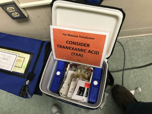
Massive Transfusion Protocol (MTP) week continues! I’m releasing the next Trauma MedEd newsletter to subscribers at the end of the week, which deals with advanced MTP topics. So leading up to that, I am reviewing the basics for the next several days. I’ll continue today with some tips on the logistics of your MTP.

MTP logistics include details such as who will be delivering the blood, what actually goes in each cooler, what ratios should be used, limitations imposed by the use of frozen plasma, and documentation. I’ll discuss details about ratios and FFP in the next Trauma MedEd newsletter.

The runners who travel between blood bank and the patient need to be selected carefully. Blood bank tech? Not ideal because they’ve got more important work to do. ED or OR tech? Maybe, as long as you’ve got a reliable pool. Student or resident? Probably not, because they may not know their way to the blood bank, which are typically placed in the farthest corner of the basement as possible. One of the most creative solutions I’ve seen is the use of a hospital security officer. Think about it. They know the hospital layout cold, including that obscure corner where the blood bank is located. And there are plenty around all the time!

Documentation is critically important, both in the trauma bay and the blood bank. Trauma activations, especially ones requiring MTP, are very fast moving and complex. Two sets of documentation are crucial: accurate records of blood product administration (on the trauma flow sheet), and documentation of just about everything else (in the blood bank). A specific timestamp on the trauma flow sheet that records the exact time of activation of MTP is a big plus.

What about coolers? I’ve seen everything used from uninsulated plastic buckets, picnic coolers, and pneumatic tube containers to large, self-contained rolling refrigeration units. The choice of container really boils down to cost vs waste. The cheaper it is, the less insulated it is, the more likely that blood products will be discarded due to high temperature if not transfused promptly. The best blend of cost vs utility seems to be the good, old-fashioned picnic cooler. It’s very portable, reasonably cheap, and can be tested for temperature maintenance. Just be sure to secure a pouch to the outside to keep platelets at room temperature to maintain full functionality.

Here’s a sample MTP cooler that’s ready for use. The platelets are in the pouch on the left. Note the reminder to prompt the team to give TXA if not contraindicated. This is often forgotten in the heat of the trauma activation.

Who actually runs your MTP? In some ways, it should run itself. Coolers get delivered, products get transfused. However, some decision making is needed to decide how long to continue and whether any tweaking of product ratios needs to happen. In the emergency department, the surgeon or emergency physician can do this. But once a trauma patient arrives in the OR, that is no longer the case. The emergency physician was left behind in the ED and the surgeon is up to her elbows in trouble. What about the anesthesiologist? Nope, they are busy keeping the patient safely asleep, regulating rapid infusions, and actually administering the blood.

Most of the time, however, these two physicians actually end up running the MTP while multi-tasking at their other job. Unfortunately, this can lead to errors and delays. One best practice to consider: a highly trained trauma resuscitation nurse or advanced practice provider (APP) can run the MTP from the sidelines. They travel with the patient from ED to OR, managing the MTP the entire time. This offloads responsibility from busier people.

Tomorrow, I’ll continue with MTP deactivation and analysis.

And don’t forget to subscribe to TraumaMed if you want to get a full newsletter discussing advanced MTP topics this Friday. Otherwise, you’ll be reading a post on CT scans and rib fractures in the elderly! Subscribe and download back issues by clicking here.

Links: (note – future links will not be live until 9am the day they are published)

Print Friendly, PDF & Email

MTP Week Part I: How To Build Your Massive Transfusion Protocol

It’s Massive Transfusion Protocol (MTP) week! I’m releasing the next Trauma MedEd newsletter to subscribers at the end of the week, which deals with advanced MTP topics. So leading up to that, I’ll be reviewing the basics for the next several days. I’ll start today with tips on how to build a solid MTP at your center.

Your massive transfusion protocol is a complex set of processes that touch many, many areas within your hospital. There are five basic components (and a few sub-components) to any MTP, so let’s dig into them one by one. They are:

  • Universality
  • Activation
  • Logistics
    • Components
    • Runners
    • Documentation
    • Coolers
  • Deactivation
  • Analysis

Let’s scrutinize each one, starting with the first two today.

Universality. This means that there should be one, and only one MTP in your hospital. I’ve seen some hospitals that have one MTP for trauma, one for cardiothoracic surgery, one for OB, one for GI, and on and on. Yes, each of those services deals with patients who are suffering from blood loss. But it’s the same blood that your trauma patients lose! There’s no need to create a protocol for each, with different ratios, extra drugs, etc. This can and will create confusion in the blood bank which may lead to serious errors.

Activation. This consists of two parts: how do we decide to activate, and then how does everyone involved find out that the MTP is actually being activated? I’ll discuss activation criteria on Thursday. But what about the notification process? Phone call? Order in the electronic medical record (EMR)? Smoke signals?

The most reliable method is a good, old-fashioned phone call. Do not use your EMR except for documentation purposes. Unless there is a very reliable system in the blood bank that translates an EMR order into an annoying alarm or flashing lights, don’t rely on this at all.

Then decide upon the minimum amount of information that the blood bank needs to begin preparing blood products. This usually consists of a name or temporary patient identifier, sex, and location of activation. Ensure that an ID or transfusion band is affixed to the patient so that wrong blood products are not given in multiple patient events.

Tomorrow, I’ll continue with the logistics of the MTP.

And don’t forget to subscribe to TraumaMed if you want to get a full newsletter discussing advanced MTP topics this Friday. Otherwise, you’ll be reading a post on CT scans and rib fractures in the elderly! Subscribe and download back issues by clicking here.

Links: (note – future links will not be live until 9am the day they are published)

Print Friendly, PDF & Email

Giving TXA Via An Intraosseous Line?

Seriously injured patients frequently develop coagulopathy, which makes resuscitation (and survival) more challenging. A few years ago, the CRASH-2 study lent support for using tranexamic acid (TXA) in select trauma patients to improve survival. This drug is cheap and has antifibrinolytic properties that may be beneficial if given for life-threatening bleeding within 3 hours of initial injury. It’s typically given as a rapid IV infusion, followed by a slower followup infusion. The US military has adopted its routine use at forward combat hospitals.

But what if you don’t have IV access? This can and does occur with military type injuries. Surgeons at Madigan Army Medical Center in Washington state tried using a common alternative access device, the intraosseous needle, to see if the results were equivalent. This study used an adult swine model with hemorrhage and aortic crossclamping to simulate military injury and resuscitation. Half of the animals then received IV TXA, the other half had it administered via IO. Only the bolus dose was given. Serum TXA levels were monitored, and serial ROTEM determinations were performed to evaluate coagulopathy.

Here are the factoids:

  • The serum TXA peak and taper curves were similar. The IV peak was higher than IO and approached statistical significance (0.053)
  • ROTEM showed that the animals were significantly hyperfibrinolytic after injury, but rapidly corrected after administration of TXA. Results were the same for both IV and IO groups.

Bottom line: This was a very simple and elegant study. The usual animal study issues come into play (small numbers, pigs are not people). But it would be nearly impossible to have such a study approved in humans. Even though the peak TXA concentration via IO is (nearly significantly) lower, this doesn’t appear to matter. The anti-fibrinolytic effect was very similar according to ROTEM analysis.

From a practical standpoint, I’m not recommending that we start giving TXA via IO in civilian practice. We don’t typically see military style injuries, and are usually able to establish some type of IV access within a reasonably short period of time. But for our military colleagues, this could be a very valuable tool!

Related posts:

Reference: No intravenous access, no problem: Intraosseous administration of tranexamic acid is as effective as intravenous in a porcine hemorrhage model. J Trauma 84(2):379-385, 2018.

Print Friendly, PDF & Email

How To Remember Those “Classes of Hemorrhage”

The Advanced Trauma Life Support course lists “classes of hemorrhage”, and various other sources list a similar classification for shock. I’ve not been able to pinpoint where these concepts came from, exactly. But I am sure of one thing: you will be tested on it at some point in your lifetime.

Here’s the table used by the ATLS course:

classes_of_shock

The question you will always be asked is:

What class of hemorrhage (or what % of blood volume loss) is the first to demonstrate systolic hypotension?

This is important because prehospital providers and those in the ED typically rely on systolic blood pressure to figure out if their patient is in trouble.

The answer is Class III, or 30-40%. But how do you remember the damn percentages?

multiscore-maxi1

It’s easy! The numbers are all tennis scores. Here’s how to remember them:

Class I up to 15% Love – 15
Class II 15-30% 15 – 30
Class III 30-40 30 – 40
Class IV >40% Game (almost) over!

Bottom line: Never miss that question again!

Print Friendly, PDF & Email

ACS Trauma Abstract #6: Scanning Unstable Trauma Patients???

If you’ve read my stuff for very long, you know I frown on sending unstable patients anywhere but to the OR. Instability tends to get worse, and that always happens at inopportune locations like hallways, elevators, and CT scanners. Imagine my surprise when I noticed an abstract being presented at the Clinical Congress of the American College of Surgeons this week suggesting that it was okay to scan hemodynamically unstable patients before “definitive therapy.”

Here’s the title:

“Computed tomography in hemodynamically abnormal thoracoabdominal trauma safely enhances surgical triage”

The devil is in the details and the language. This group from USC included all patients who were hemodynamically abnormal on arrival to the trauma bay but who normalized to SBP > 90 during the resuscitation were included.  A total of 253 of these patients were reviewed over a 9 year period, and the usual variables were analyzed (mortality, complications, hospital, ICU, and vent days, etc).

Here are the factoids:

  • Of the 253 patients studied, 45 went to straight to OR and 208 were taken to CT
  • Injury severity was identical for the two groups
  • Lengths of stay and mortality were not different, but only p values were given
  • Patients taken to CT cleared their lactic acidosis faster (12 vs 5 hours), and used a bit less plasma and significantly less blood transfusions
  • The OR group underwent more procedures (31% vs 13%), although what these were and when they were performed is not listed

Bottom line: The title of this abstract is misleading, and may fool those who don’t read the rest of the abstract. It should read:

“Computed tomography in previously hemodynamically abnormal thoracoabdominal trauma safely enhances surgical triage”

Someone who just skims through this issue of the journal may get the idea that it’s okay to scan an unstable patient. The authors are not saying this at all. If you read the conclusion carefully, you can see that the patients had to be resuscitated to a SBP > 90 before they considered taking to scan. And they did that for the majority of these patients.

The real question is, why do the scanned patients clear their lactic acidosis faster, need less blood, and undergo fewer procedures? It appears that there is some bias or selection process in play. Otherwise, why not use the magic CT scanner to make them all better?

Reference: Computed tomography in hemodynamically abnormal thoracoabdominal trauma safely enhances surgical triage. JACS 225(4S2):e175-176, 2017.

Print Friendly, PDF & Email