Category Archives: Imaging

The Lead Gown Pull-Up?

Trauma Team members typically wear a lead gown under their personal protective equipment so they don’t have to run out of the room when x-rays are taken. How often do you see people do this?

Is it really necessary? Or is it just a way to exercise your pecs and biceps? Tomorrow I’ll talk about how much radiation team members are really exposed to so you can decide if this is really necessary.

Print Friendly, PDF & Email

Contrast Extravasation Into The Psoas Muscle

Contrast extravasation after major trauma can be very problematic. Extravasation into a solid organ (liver, spleen) generally requires a quick trip to interventional radiology or the operating room. Bleeding from the bowel mesentery assures an exploratory laparotomy. Gluteal vessel extravasation is best treated with angioembolization.

But what about extravasation from off the beaten path areas like the psoas muscle? This is an uncommon finding on trauma CT, so less is known about the usual clinical course. A group in Okayama Japan performed a 10-year retrospective review of data from their hospital. They reviewed hematoma size, associated injuries, and the relationship to treatment options.

Here are the factoids:

  • 762 contrast CTs were performed due to blunt trauma over the 10 year period (only 76 per year?!)
  • About 15% (117 patients) had either lumbar process fracture or psoas hematoma, and about one quarter had obvious contrast extravasation into the muscle
  • Patients with contrast extravasation were significantly older, had higher ISS, and were more likely to require transfusion
  • There was an association between the number of transverse process fractures and “need for” angioembolization
  • Size of the psoas hematoma was predictive of the need for angioembolization
  • Angioembolization of the psoas was frequently associated with  embolization of the pelvis

The right psoas has both contrast extravasation and a sizable hematoma

Bottom line: This study has many weaknesses, but does show that psoas extravasation occurs somewhat frequently, even at a low volume center. I always worry about studies that state something like “and xx patients required intervention.” Generally, this means that it was performed at the discretion of the clinician and no consistent rules were applied. And even though hematoma size was significantly correlated with angioembolization, it’s probably not worth the effort to have your radiologist calculate it. But it does illustrate one nearly universal trauma rule:

Patients with active extravasation on CT are bleeding to death until proven otherwise

Do not sit back and manage expectantly! The corollary to this rule is:

Contrast extravasation on CT always requires active measures to stop it

These active measures are typically angioembolization for difficult to reach areas in hemodynamically stable patients (gluteal artery for buttock, lumbar artery for psoas muscle, solid organs). Unstable patients absolutely require a trip to the OR for control. Superficial muscular bleeding frequently stops with good pressure dressings or positioning the patient so they lie on the affected area. Just don’t sit around and watch these patients bleed when you see extravasation on the CT.

Reference: Impact of contrast extravasation on computed tomography of thepsoas major muscle in patients with blunt torso trauma. J Trauma 86(2):268-273, 2019.

Print Friendly, PDF & Email

EAST 2019 #10: Incidental Findings In Trauma Imaging

Every major trauma patient undergoes some type of radiographic imaging during their initial evaluation. On occasion, some incidental finding unrelated to trauma shows up unexpectedly. These incidentalomas add several additional layers of complexity to the evaluation process.

What does the finding mean? Is it important? How do I tell the patient? Their primary care provider? When? Many times, these findings have little clinical significance. But on occasion, they can be life changing, such as the incidental renal cell carcinoma.

The group at University of Tennessee – Knoxville reviewed one year of incidental findings in trauma evaluations at their Level I trauma center. They specifically looked at diagnoses with malignant potential, and how findings were disclosed to the patient.

Here are the factoids:

  • Over 6000 patients were reviewed, and 22% had 1222 incidental findings (that’s 2 per patient!)
  • The findings were noted in males about 2/3 of the time
  • 59% of of incidentalomas were in the chest, and 16% in the abdomen
  • The most common findings were lung nodule (209), hernia (112), and renal cyst (103)
  • Only 60% of patients were informed prior to discharge (!)
  • Trauma registry abstraction resulted in an additional 20% of patients informed of the finding
  • 58 patients could not be located, and in 43 patients there was no documented attempt to contact them
  • An additional 100 registry charts that did not contain incidental findings were re-abstracted and searched for incidental findings. Nearly one third contained incidental findings!
  • If the incidental finding was noted in the radiology report summary, 78% of patients were informed. But when it was buried in the body of the report, only 22% were disclosed.

Here are some questions for the authors and presenter to consider in advance to help them prepare for audience questions:

  • The majority of the incidental findings were in the chest and abdomen. What and where were the rest?
  • What would you recommend for achieving optimal disclosure based on your results? It appears that 20% or so of patients never learned of the finding.
  • What should we do about our registry data? Should we force our registrars to comb all reports for possible incidental findings? Given that one fifth of patients have them (or more) that seems like a lot of work!
  • How has your work changed your practice at UT Knoxville?

This is a fascinating paper, and gives me some ideas for upcoming blog posts! I will definitely be in the audience for this presentation.

Reference:  A novel use of the trauma registry: incidental findings in the trauma patient. EAST 2019, Quick Shot Paper #13.

Print Friendly, PDF & Email

Radiation Exposure From Imaging At Adult vs Pediatric Trauma Centers

Anyone who reads this blog already knows I am a big believer in well-crafted and focused practice guidelines. And by focused I mean directed toward a clinical problem that typically sees a lot of variability between care providers. Use of imaging is one of these clinical problems. A surgeon may order a certain set of studies for a major blunt trauma patient, and their emergency medicine colleague might order a somewhat different set for someone with the exact same history, physical exam, and injury pattern. Who is right? Neither!

And the variability is even greater when we throw a pediatric patient into the mix. Trauma professionals tend to be even more “generous” when ordering studies on children because they are afraid they might miss something. Unfortunately, this has the potential for overuse of imaging and exposure to unnecessary radiation.

Avery Nathens and a consortium of pediatric trauma centers used the Trauma Quality Improvement Database (TQIP) to review CT imaging practices on children age < 18 over a four year period. Only blunt trauma patients were studied, and the Abbreviated Injury Scale had to be at least 2 for a minimum of one organ system. Transfer patients were excluded because there is no data on imaging for the referring hospital in the TQIP database for them. Comparisons were made between practices at adult trauma centers treating children (ATC), mixed adult/pediatric centers (MTC) and pediatric only trauma centers (PCT).

Here are the factoids:

  • Over 59,000 pediatric trauma patients were identified in the data, and about half (31,081) received at least one CT scan
  • The distribution among the three types of trauma centers was even, with roughly a third seen at each
  • Of the study group 46% had a head CT, 17% a chest CT, and 26% underwent abdominal CT
  • Injured children were more likely to undergo CT if they were older, had a higher ISS, lower motor GCS, were involved in a car crash, or had severe injuries to head or torso
  • Overall CT rates were about the same across the three types of centers (56% ATC, 57% MTC, 43% PTC)
  • Chest CT was performed 8x as much at ATC/MTC vs PTC (!)
  • Abdominal CT was performed 2x as much at ATC/MTC vs PTC
  • Lesser injured children received relatively more CT scans at ATC/MTC when compared to PTC
  • Using standard estimates of cancer risk from all CT scans received, children treated at adult or mixed trauma centers received enough radiation to cause 17 additional lifetime cancers per 100,000 patients
  • About 35 additional lifetime cancers per 100,000 would be caused by the chest and abdominal scans performed at the ATC/MTC centers when compared to pediatric-only centers

Bottom line: This is yet another reason to adopt a well-designed pediatric imaging guideline. Not only are adult centers using CT scanning much more that pediatric-only centers, but they are unnecessarily adding to the lifetime risk for cancer of our children!

As I always recommend, find a well-designed imaging guideline from an established pediatric center and “borrow” it. Sure, it may need a few minor tweaks to fit well with your hospital. That’s okay. Just get it done so your team can begin to order the initial imaging studies consistently and intelligently.

Reference: Computed tomography rates and estimated radiation-associated cancer risk among injured children treated at different trauma center types. Injury 2018, in press.  https://doi.org/10.1016/j.injury.2018.09.036

Print Friendly, PDF & Email

Pan Scanning for Elderly Falls?

The last abstract for the Clinical Congress of the American College of Surgeons that I will review deals with doing a so-called “pan-scan” for ground level falls. Apparently, patients at this center have been pan-scanned for years, and they wanted to determine if it was appropriate.

This was a retrospective trauma registry review of 9 years worth of ground level falls. Patients were divided into young (18-54 years) and old (55+ years) groups. They were included in the study if they received a pan-scan.

Here are the factoids:

  • Hospital admission rates (95%) and ICU admission rates (48%) were the same for young and old
  • ISS was a little higher in the older group (9 vs 12)
  • Here are the incidence and type of injuries detected:
Young (n=328) Old (n=257)
TBI 35% 40%
C-spine 2% 2%
Blunt Cereb-vasc inj * 20% 31%
Pneumothorax 14% 15%
Abdominal injury 4% 2%
Mortality * 3% 11%

 * = statistically significant

Bottom line: There is an ongoing argument, still, regarding pan-scan vs selective scanning. The pan-scanners argue that the increased risk (much of which is delayed or intangible) is worth the extra information. This study shows that the authors did not find much difference in injury diagnosis in young vs elderly patients, with the exception of blunt cerebrovascular injury.

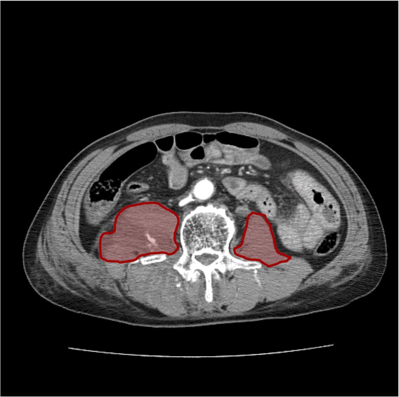
Most elderly patients who fall sustain injuries to the head, spine (all of it), extremities and hips. The torso is largely spared, with the exception of ribs. In my opinion, chest CT is only for identification of aortic injury, which just can’t happen from falling over. Or even down stairs. And solid organ injury is also rare in this group.

Although the future risk from radiation in an elderly patient is probably low, the risk from the IV contrast needed to see the aorta or solid organs is significant in this group. And keep in mind the dangers of screening for a low probability diagnosis. You may find something that prompts invasive and potentially more dangerous investigations of something that may never have caused a problem!

I recommend selective scanning of the head and cervical spine (if not clinically clearable), and selective conventional imaging of any other suspicious areas. If additional detail of the thoracic and/or lumbar spine are needed, specific spine CT imaging should be used without contrast.

Related posts:

Reference: Pan-scanning for ground level falls in the elderly: really? ACS Surgical Forum, trauma abstracts, 2016.

Print Friendly, PDF & Email